Clinical Echocardiography
-
Introduction to echocardiography and ultrasound imaging12 Chapters
-
Physics of ultrasound
-
The ultrasound transducer
-
Technical aspects of the ultrasound image
-
Two-dimensional (2D) echocardiography
-
Optimization of the ultrasound image
-
M-mode (motion mode) echocardiography
-
Doppler effect and Doppler echocardiography
-
Pulsed Wave Doppler
-
Continuous Wave Doppler (CW Doppler)
-
Color Doppler
-
Tissue Doppler (Tissue Velocity Imaging)
-
Artifacts in ultrasound imaging
-
Physics of ultrasound
-
Principles of hemodynamics5 Chapters
-
The echocardiographic examination3 Chapters
-
Left ventricular systolic function and contractility11 Chapters
-
Left Ventricular Function
-
Myocardial Mechanics: Structure and Function of Myocardial Fibers
-
Ventricular Pressure-Volume Relationship: Preload, Afterload, Stroke Volume, Wall Stress & Frank-Starling's law
-
Assessing left ventricular systolic function
-
Left ventricular mass and volume (size)
-
Ejection fraction (EF): Physiology, Measurement & Clinical Evaluation
-
Fractional shortening for estimation of ejection fraction
-
Strain, strain rate and speckle tracking: Myocardial deformation
-
Left Ventricular Segments for Echocardiography and Cardiac Imaging
-
The Coronary Arteries
-
Regional Myocardial Contractile Function: Wall Motion Abnormalities
-
Left Ventricular Function
-
Left ventricular diastolic function3 Chapters
-
Cardiomyopathies6 Chapters
-
Heart failure: Causes, types, diagnosis, treatments & management
-
Echocardiography in cardiomyopathies: an overview
-
Hypertrophic Cardiomyopathy (HCM) & Hypertrophic Obstructive Cardiomyopathy (HOCM)
-
Dilated Cardiomyopathy (DCM): Definition, Types, Diagnostics & Treatment
-
Arrhythmogenic Right Ventricular Cardiomyopathy / Dysplasia (ARVC, ARVD)
-
Tachycardia induced cardiomyopathy
-
Heart failure: Causes, types, diagnosis, treatments & management
-
Valvular heart disease8 Chapters
-
Miscellaneous conditions5 Chapters
-
Pericardial disease2 Chapters
Left ventricular mass and volume (size)
Size and mass of the left ventricle
Left ventricular mass is a flawed proxy for ventricular systolic function and load. The mass is, however, an important parameter in the assessment of ventricular hypertrophy and cardiomyopathy. Numerous formulas have been developed to approximate ventricular mass. Most formulas are simple mathematical equations, included in all ultrasound systems, and assume that the geometry of the left ventricle is normal.
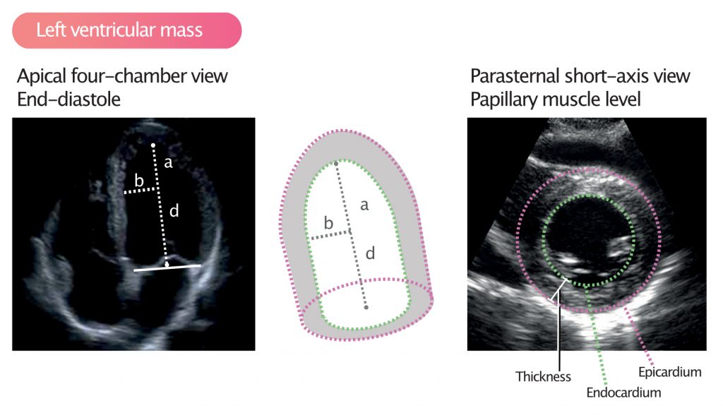
massLV = 1.05 (masstotal – masscavity)
LV = left ventricle; 1.05 = mycoardial mass constant.
Left ventricular hypertrophy (LVH)
A diagnosis of left ventricular hypertrophy is based on total left ventricular mass, which can be calculated by obtaining the measurements shown in Figure 1. The ultrasound system automatically calculates RWT (Relative Weight Thickness), provided that the patient’s weight, height and sex are entered. RWT is a measure of the type of hypertrophy.
Generally, hypertrophy is defined as wall thickness exceeding 12 mm. Thus, wall thickness >12 mm should raise suspicion of hypertrophy.

