Clinical Echocardiography
-
Introduction to echocardiography and ultrasound imaging12 Chapters
-
Physics of ultrasound
-
The ultrasound transducer
-
Technical aspects of the ultrasound image
-
Two-dimensional (2D) echocardiography
-
Optimization of the ultrasound image
-
M-mode (motion mode) echocardiography
-
Doppler effect and Doppler echocardiography
-
Pulsed Wave Doppler
-
Continuous Wave Doppler (CW Doppler)
-
Color Doppler
-
Tissue Doppler (Tissue Velocity Imaging)
-
Artifacts in ultrasound imaging
-
Physics of ultrasound
-
Principles of hemodynamics5 Chapters
-
The echocardiographic examination3 Chapters
-
Left ventricular systolic function and contractility11 Chapters
-
Left Ventricular Function
-
Myocardial Mechanics: Structure and Function of Myocardial Fibers
-
Ventricular Pressure-Volume Relationship: Preload, Afterload, Stroke Volume, Wall Stress & Frank-Starling's law
-
Assessing left ventricular systolic function
-
Left ventricular mass and volume (size)
-
Ejection fraction (EF): Physiology, Measurement & Clinical Evaluation
-
Fractional shortening for estimation of ejection fraction
-
Strain, strain rate and speckle tracking: Myocardial deformation
-
Left Ventricular Segments for Echocardiography and Cardiac Imaging
-
The Coronary Arteries
-
Regional Myocardial Contractile Function: Wall Motion Abnormalities
-
Left Ventricular Function
-
Left ventricular diastolic function3 Chapters
-
Cardiomyopathies6 Chapters
-
Heart failure: Causes, types, diagnosis, treatments & management
-
Echocardiography in cardiomyopathies: an overview
-
Hypertrophic Cardiomyopathy (HCM) & Hypertrophic Obstructive Cardiomyopathy (HOCM)
-
Dilated Cardiomyopathy (DCM): Definition, Types, Diagnostics & Treatment
-
Arrhythmogenic Right Ventricular Cardiomyopathy / Dysplasia (ARVC, ARVD)
-
Tachycardia induced cardiomyopathy
-
Heart failure: Causes, types, diagnosis, treatments & management
-
Valvular heart disease8 Chapters
-
Miscellaneous conditions5 Chapters
-
Pericardial disease2 Chapters
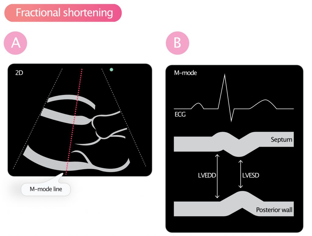
Fractional shortening for estimation of ejection fraction
Fractional shortening (FS) for estimating systolic function
Fractional shortening (FS) is calculated by measuring the percentage change in left ventricular diameter during systole. It is measured in parasternal long axis view (PLAX) using M-mode. The end-systolic and end-diastolic left ventricular diameters are measured. The following formula is used to calculate fractional shortening:
FS (%) = (LVEDD – LVESD / LVEDD) • 100
Fractional shortening is a rather poor measure of left ventricular systolic function. This is due to the following reasons:
- Left ventricular geometry must be normal.
- There must not be regional differences in contractile function. Otherwise, the point of measurement may not be representative.
- Ventricular activation must be normal. For example, in the setting of left bundle branch block (LBBB), fractional shortening is not representative of ventricular function, since the activation proceeds abnormally.
Normal value for fractional shortening (FS)
| Normal FS, M-mode | >25% |
| Normal FS, 2D measurement | >18% |
Advantages of fractional shortening
If ventricular geometry is normal and there are no regional wall motion abnormalities, then fractional shortening correlates strongly with ejection fraction. Similar to ejection fraction, fractional shortening is affected by preload and afterload. It possible to calculate fractional shortening using measurements in 2D.

