Hemodynamic calculations with PISA (Proximal Isovelocity Surface Area)
PISA (Proximal Isovelocity Surface Area) is a phenomenon that occurs when liquid flows through a circular orifice. The flow will converge and accelerate just proximal to the orifice. The change in flow profile results in the formation of a hemisphere with several layers. Flow velocity is equal within each layer (Figure 1).
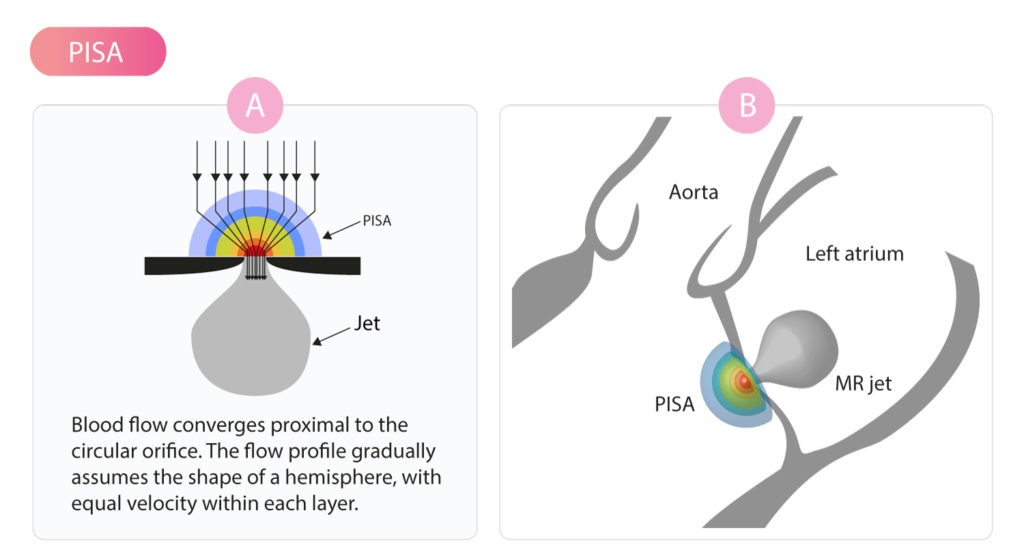
PISA is the hemisphere itself. It appears as a semicircle in 2D images (Figure 1). The radius of PISA can be used to calculate the diameter of the orifice. This has fundamental clinical implications as it enables the investigator to calculate the area of stenoses and regurgitations. Such area estimations are fundamental in the management of valvular conditions, such as aortic stenosis, aortic regurgitation, mitral valve stenosis, mitral valve regurgitation, etc. The radius of PISA is measured from the surface of the hemisphere to the narrowest segment of the Doppler beam, which is located within the orifice (Figure 2).
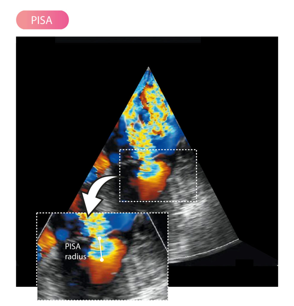
Color Doppler is used to revealing PISA. As discussed earlier, aliasing occurs when using color Doppler to analyze velocities greater than the Nyquist limit. Aliasing implies that neither the direction nor velocity of flow can be determined. This results in the Doppler signal shifting color, such that blue turns red, and red turns blue. For color Doppler aliasing usually occurs when velocities exceed 0.5 m/s, which they generally do in the setting of significant stenoses and regurgitations.
Thus, aliasing is exploited to reveal PISA. An optimal assessment of PISA requires adjusting the Nyquist limit until PISA assumes the shape of a semicircle. The radius and area of PISA are calculated as follows:
areaPISA = 2 • π • rPISA2
The flow (Q) can be calculated using PISA, as follows:
QPISA = areaPISA • valiasing
valiasing = aliasing speed
According to the principle of continuity, the flow in PISA must be equivalent to the flow through the orifice itself. This implies that PISA can be used to quantify regurgitation volume. In the case of mitral regurgitation (MR), the regurgitant area can be calculated using the following formula:
areaMR = 2 • π • rPISA • (valiasing / VmaxMR)
MR = mitral regurgitation; VmaxMR = maximum velocity of mitral regurgitation; valiasing = aliasing speed.
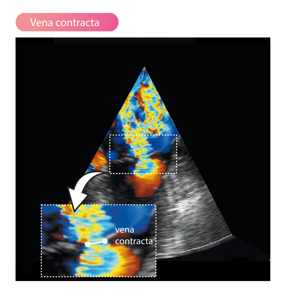
This formula actually calculates the area of vena contracta (Figure 3), which is approximately equal to the area of the orifice. The areaMR is also called EROA (Effective Regurgitant Orifice Area).
The regurgitant volume (RV) can be calculated by the following formula:
RV = areaMR • VTIMR
RV = regurgitant volume; VTI = velocity time integral.
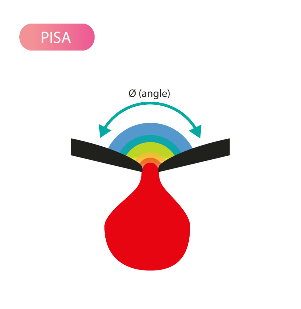
These formulas for PISA performs best when the surface surrounding the orifice is flat, which is often not the case for the valves. For example, a closed aortic valve assumes the shape of a cone. Fortunately, this can be accounted for by including a correction for the angle, as follows:
areaPISA = 2 • π • rPISA2 • (Ø / 180)
Ø = angle.
Figure 4 shows the angle to be measured.
The width of vena contracta can also be used to estimate the severity of a regurgitation.

