Principles of image optimization in echocardiography
In order to obtain optimal ultrasound images, it is necessary to adjust several parameters continuously during the examination. Typically, examinations in each echocardiographic view (also called window) are initialized by identifying an overview image. Starting in the overview image the depth is reduced as much as possible. Reducing the depth results in increased frame rate and thus better image resolution. If possible, the width of the image is also reduced, which likewise results in increased image-resolution. It is also possible to zoom in on regions of interest; e.g the aortic valve can be zoomed in to study its anatomy and function. Zooming in improves the resolution in a particular area. Alternatively, it is possible to place the focus at the level of the region of interest. The difference between zooming in and shifting focus is that the zoom encloses a specific region of the image, whereas shifting focus simply adjusts the location (along the ultrasound beam) with the best resolution. If the ultrasound image is too dark, it is possible to increase the gain. This amplifies the incoming (reflected) ultrasound waves such that each object appears whiter on the image. Increasing gain excessively results in lower resolution and difficulties discerning tissue borders. These are the main adjustments made to improve image quality.
Adjusting image depth and zoom
Examinations in each echocardiographic view are initialized by identifying an overview image. Starting in the overview image the depth is reduced as much as possible, without excluding regions of interest. Reducing the depth results in an increased frame rate and thus enhanced image resolution. If a particular region is of interest, that region can be zoomed in. Note that the image becomes more grainy as the zoom is increased.
Gain: signal amplification
The ultrasound machine amplifies all incoming (reflected) ultrasound waves. However, the examiner can further increase the amount of gain applied to incoming sound waves. This is done using gain control or time-gain compensation (TGC).

Gain control: overall gain
Gain control regulates the global (overall) gain. Increasing the overall gain will increase the gain for all reflected sound waves, making all objects in the image whiter. This may clarify some tissue borders but excessive use of gain results in deterioration of image quality.
Time gain compensation / control (TGC)
Time-gain control / compensation (TGC) adjusts the gain at specific levels along the ultrasound field. The purpose of TGC is to gradually increase the amount of gain as the depth increases; this compensates for the attenuation that occurs with increasing depth. TGC is adjusted using multiple controls that each represent a specific depth in the image (Figure 1). The bottom control adjusts gain at the bottom of the image etc. TGC is generally increased at the bottom of the image since the ultrasound lines have the lowest density there (and thus the lowest image resolution). TGC at the top of the image is usually kept at low levels.
Frequency of ultrasound waves
Low ultrasound wave frequency provides high tissue penetration and low image resolution. High-frequency waves provide good image resolution but worse penetration. Visualizing objects located in close proximity to the transducer, therefore, is done using high-frequency waves. The frequency of the ultrasound wave must generally be reduced in order to visualize objects located far away from the transducer. Hence, using low-frequency waves to visualize distant objects is motivated by the advantages of greater tissue penetration of such waves.
Recommended: Physics of Ultrasound
Image focus
The focus is placed at the level where the one examines is located. One can choose to place one or more focus (multiple focus lowers the image update rate).
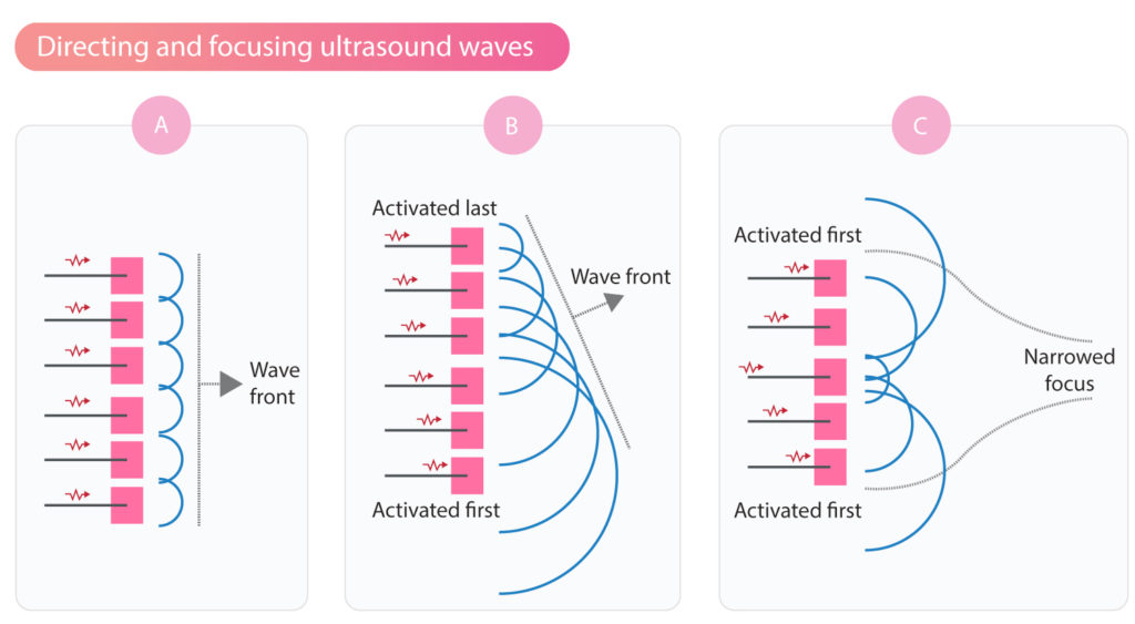
The direction and focus of ultrasound waves can be adjusted by varying the sequence of activation of the piezoelectric crystals (Figure 2). If the activation starts at the lateral crystals and proceeds towards the center, then the ultrasound beam will be focused (Figure 2C). The focus can be placed anywhere along the ultrasound field.
Frame rate
Temporal image resolution is the ability to describe the movement of objects over time. Echocardiography requires high temporal resolution to study the detailed movements of relatively small objects. In order to produce recordings with high temporal resolution, it is critical to produce images rapidly. The more images that can be produced and presented per unit of time (i.e frame rate), the greater the temporal resolution.
It is possible to manually increase the frame rate (of any obtained image) to a certain extent. Frame rate is always increased when reducing the width and depth of the ultrasound image.

