Left Ventricular Segments for Echocardiography and Cardiac Imaging
Standardized myocardial segmentation for echocardiography and cardiac imaging
Cardiovascular imaging modalities have developed rapidly in the past few decades. The repertoire of available methods includes echocardiography, cardiovascular magnetic resonance (CMR), cardiac computed tomography (CT), single-photon emission computed tomography (SPECT), positron emission computed tomography (PET) and coronary angiography. These modalities aim to image the myocardium, assess wall motions and myocardial perfusion. However, due to varying strengths, limitations and clinical applications of these modalities, the definitions of cardiac planes, ventricular segments, and coronary arterial territories evolved differently. This resulted in difficulties when comparing examinations in clinical practice and research. Therefore, the American Heart Association (AHA) and the European Society for Cardiology (ESC) have established a standardization for ventricular segmentation, nomenclature and assignment of coronary arterial territory (Cerqueira et al).
The purpose of standardizing ventricular segmentation, arterial territories and nomenclature is to develop consistency across examinations. This is of utmost importance when assessing myocardial structure, perfusion and wall motion.
Cardiac planes
All imaging modalities define, orient and display the heart using the long axis of the left ventricle and selected planes oriented at 90° angles relative to the long axis. With regards to echocardiography, this implies that the following planes are used: short axis, vertical long axis, and horizontal long axis. These three planes correspond to the parasternal short-axis view (PSAX), apical two-chamber view (A2C), and apical four-chamber view (A4C), respectively (Table 1).
Table 1. Cardiac Planes in Echocardiography
| Plane | Echocardiographic view |
| Short axis | Parasternal short-axis view (PSAX) |
| Vertical long axis | Apical two-chamber view (A2C) |
| Horizontal long axis | Apical four-chamber view (A4C) |
Segments of the left ventricle
Based on anatomical landmarks and autopsy studies (Edwards et al), the left ventricle is divided into three equal parts along the long axis of the ventricle. This creates three circular sections of the left ventricle named basal, mid-cavity, and apical. These three parts are divided into a total of 17 segments (Figure 1). Six basal segments, constituting 35% of myocardial mass; six mid-cavity segments, constituting 35% of myocardial mass and 5 apical segments, constituting 30% of myocardial mass. The 17-segment model provides the best agreement with anatomic data, as compared with other segmentation models.
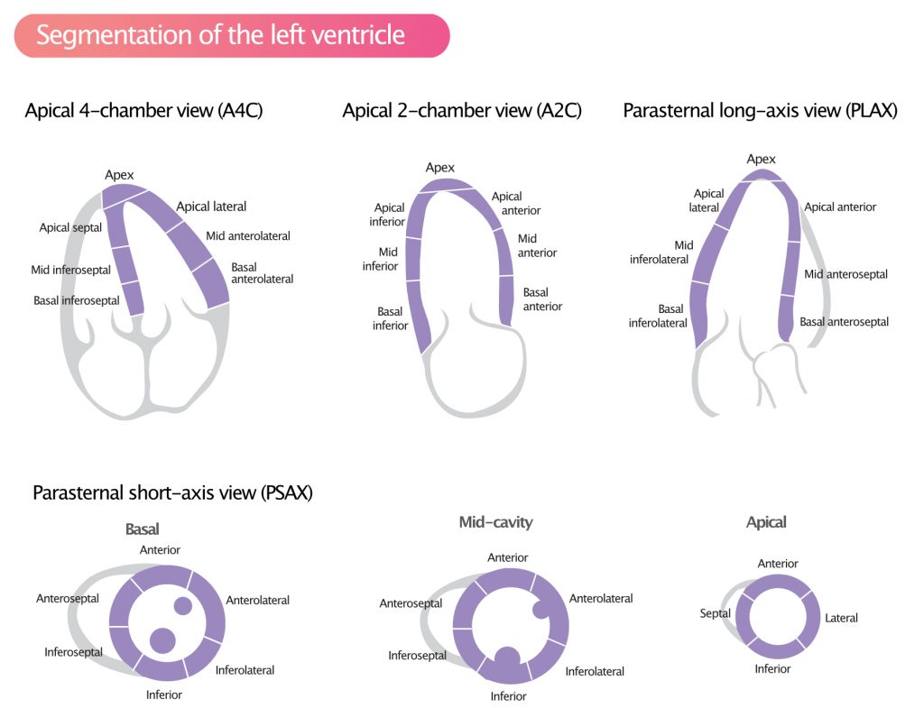
The basal part is divided into six segments of 60° each. The segments along the circumference are basal anterior, basal anteroseptal, basal inferoseptal, basal inferior, basal inferolateral, and basal anterolateral. The mid-cavity part is also divided into six 60° segments (mid anterior, mid anteroseptal, mid inferoseptal, mid inferior, mid inferolateral, and mid anterolateral). The apical part is divided into four 90° segments (apical anterior, apical septal, apical inferior, and apical lateral) and the apex (Table 2).
Table 2. The 17 segments of the left ventricle
| Basal Segments | Mid-cavity Segments | Apical Segments | |||
| 1. | basal anterior | 7. | mid anterior | 13. | apical anterior |
| 2. | basal anteroseptal | 8. | mid anteroseptal | 14. | apical septal |
| 3. | basal inferoseptal | 9. | mid inferoseptal | 15. | apical inferior |
| 4. | basal inferior | 10. | mid inferior | 16. | apical lateral |
| 5. | basal inferolateral | 11. | mid inferolateral | 17. | apex |
| 6. | basal anterolateral | 12. | mid anterolateral |
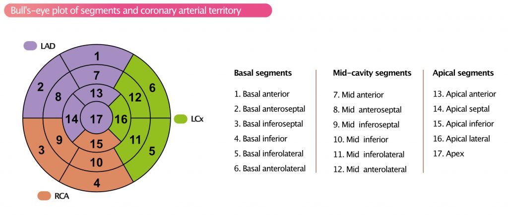
These 17 segments can be arranged as a polar (bull’s-eye) plot with the apex in the center, the four apical segments as the first ring, the six mid-cavity segments as the second ring, and the six basal segments as the outer ring (Figure 2).
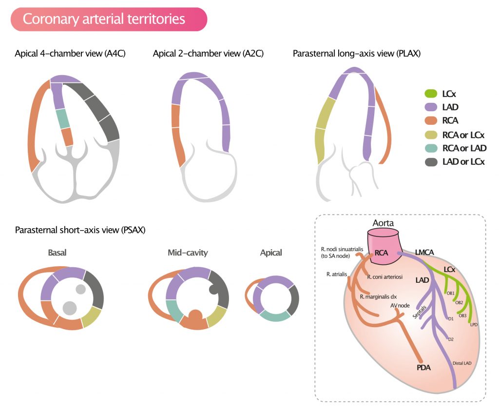
The attachment of the right ventricular wall to the left ventricle is used to identify and separate the septum from the left ventricular anterior and inferior free walls.
References
Edwards et al: Standardized nomenclature and anatomic basis for regional tomographic analysis of the heart. Mayo Clin Proc. 1981;56:479–497.

