Clinical Echocardiography
-
Introduction to echocardiography and ultrasound imaging12 Chapters
-
Physics of ultrasound
-
The ultrasound transducer
-
Technical aspects of the ultrasound image
-
Two-dimensional (2D) echocardiography
-
Optimization of the ultrasound image
-
M-mode (motion mode) echocardiography
-
Doppler effect and Doppler echocardiography
-
Pulsed Wave Doppler
-
Continuous Wave Doppler (CW Doppler)
-
Color Doppler
-
Tissue Doppler (Tissue Velocity Imaging)
-
Artifacts in ultrasound imaging
-
Physics of ultrasound
-
Principles of hemodynamics5 Chapters
-
The echocardiographic examination3 Chapters
-
Left ventricular systolic function and contractility11 Chapters
-
Left Ventricular Function
-
Myocardial Mechanics: Structure and Function of Myocardial Fibers
-
Ventricular Pressure-Volume Relationship: Preload, Afterload, Stroke Volume, Wall Stress & Frank-Starling's law
-
Assessing left ventricular systolic function
-
Left ventricular mass and volume (size)
-
Ejection fraction (EF): Physiology, Measurement & Clinical Evaluation
-
Fractional shortening for estimation of ejection fraction
-
Strain, strain rate and speckle tracking: Myocardial deformation
-
Left Ventricular Segments for Echocardiography and Cardiac Imaging
-
The Coronary Arteries
-
Regional Myocardial Contractile Function: Wall Motion Abnormalities
-
Left Ventricular Function
-
Left ventricular diastolic function3 Chapters
-
Cardiomyopathies6 Chapters
-
Heart failure: Causes, types, diagnosis, treatments & management
-
Echocardiography in cardiomyopathies: an overview
-
Hypertrophic Cardiomyopathy (HCM) & Hypertrophic Obstructive Cardiomyopathy (HOCM)
-
Dilated Cardiomyopathy (DCM): Definition, Types, Diagnostics & Treatment
-
Arrhythmogenic Right Ventricular Cardiomyopathy / Dysplasia (ARVC, ARVD)
-
Tachycardia induced cardiomyopathy
-
Heart failure: Causes, types, diagnosis, treatments & management
-
Valvular heart disease8 Chapters
-
Miscellaneous conditions5 Chapters
-
Pericardial disease2 Chapters
Tissue Doppler (Tissue Velocity Imaging)
Tissue Doppler (Tissue Velocity Imaging)
Previous chapters on Doppler imaging have all focused on measurements of blood flow. However, the Doppler effect can also be used to study myocardial motion. Myocardial motion during systole and diastole alter the frequency of ultrasound waves that are reflected back to the transducer. There are two fundamental differences between blood and myocardium in terms of the reflected ultrasound waves. (1) The velocity of myocardial motion is significantly lower than the velocity of blood flow. Therefore, ultrasound waves reflected from myocardium will have a lower Doppler shift, as compared with waves reflected from erythrocytes. (2) Whereas reflections from erythrocytes have low amplitude, sound waves reflected from myocardium have high amplitude. This is due to the high density of myocardium. It follows that myocardium generates reflections with high amplitude and low Doppler shift. To analyze reflections from the myocardium, the ultrasound machine filters out all other reflected sound waves, such that only those representing myocardium are recorded.
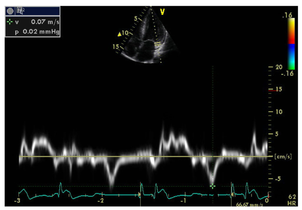
Pulsed tissue Doppler
Pulsed tissue Doppler uses a sample volume where the velocity is recorded (Figure 1).
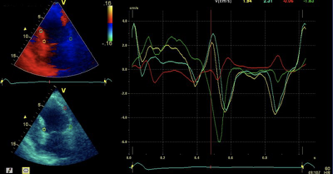
Color Tissue Doppler
Color tissue Doppler analyzes myocardial velocities within a color sector. Myocardium moving towards the transducer is colored red, and myocardium moving away from the transducer is colored blue. The advantage of color tissue Doppler is that all myocardium is analyzed simultaneously, which makes it possible to compare myocardial regions (Figure 2).
It is important to note that the apex of the heart is fixed to the diaphragm via pericardium and connective tissue. Therefore the apex does not move much during the cardiac cycle, despite the fact that cells in the apex contract as much as the cells in the basal parts. Since the apex is fixed and there are longitudinal muscle fibers stretching from the apex to the basal parts, it appears as if basal regions are pulled down towards the apex. The longitudinal muscle fibers generate the longitudinal contraction (or longitudinal shortening) during systole.
It is also important to note that the velocity recorded in one region does not depend on the function in the region, but rather the function (contractility) of all myocardium located apically to the point of measurement.
Tissue Tracking
Tissue tracking is used to calculate the distance that the myocardium moves during the cardiac cycle. The predominant technique for tissue tracking is speckle tracking, which is discussed later.

