Clinical Echocardiography
-
Introduction to echocardiography and ultrasound imaging12 Chapters
-
Physics of ultrasound
-
The ultrasound transducer
-
Technical aspects of the ultrasound image
-
Two-dimensional (2D) echocardiography
-
Optimization of the ultrasound image
-
M-mode (motion mode) echocardiography
-
Doppler effect and Doppler echocardiography
-
Pulsed Wave Doppler
-
Continuous Wave Doppler (CW Doppler)
-
Color Doppler
-
Tissue Doppler (Tissue Velocity Imaging)
-
Artifacts in ultrasound imaging
-
Physics of ultrasound
-
Principles of hemodynamics5 Chapters
-
The echocardiographic examination3 Chapters
-
Left ventricular systolic function and contractility11 Chapters
-
Left Ventricular Function
-
Myocardial Mechanics: Structure and Function of Myocardial Fibers
-
Ventricular Pressure-Volume Relationship: Preload, Afterload, Stroke Volume, Wall Stress & Frank-Starling's law
-
Assessing left ventricular systolic function
-
Left ventricular mass and volume (size)
-
Ejection fraction (EF): Physiology, Measurement & Clinical Evaluation
-
Fractional shortening for estimation of ejection fraction
-
Strain, strain rate and speckle tracking: Myocardial deformation
-
Left Ventricular Segments for Echocardiography and Cardiac Imaging
-
The Coronary Arteries
-
Regional Myocardial Contractile Function: Wall Motion Abnormalities
-
Left Ventricular Function
-
Left ventricular diastolic function3 Chapters
-
Cardiomyopathies6 Chapters
-
Heart failure: Causes, types, diagnosis, treatments & management
-
Echocardiography in cardiomyopathies: an overview
-
Hypertrophic Cardiomyopathy (HCM) & Hypertrophic Obstructive Cardiomyopathy (HOCM)
-
Dilated Cardiomyopathy (DCM): Definition, Types, Diagnostics & Treatment
-
Arrhythmogenic Right Ventricular Cardiomyopathy / Dysplasia (ARVC, ARVD)
-
Tachycardia induced cardiomyopathy
-
Heart failure: Causes, types, diagnosis, treatments & management
-
Valvular heart disease8 Chapters
-
Miscellaneous conditions5 Chapters
-
Pericardial disease2 Chapters
Continuous Wave Doppler (CW Doppler)
Continuous Wave Doppler
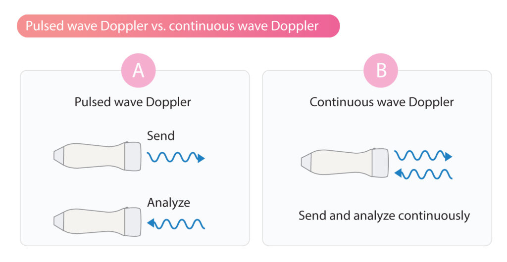
In continuous wave Doppler (CW Doppler), ultrasound waves are continuously emitted from the transducer and the reflections of these waves are analyzed continuously (Figure 1). This is possible by using two different sets of piezoelectric crystals; one set for sending ultrasound and the other for analyzing reflected sound waves. The cardinal difference between continuous wave Doppler and pulsed wave Doppler is that ultrasound is emitted and analyzed continuously in the former. This allows much higher velocitites to be measured. Continuous wave Doppler is therefore not limited by pulse repetition frequency (PRF).
The disadvantage of continuous wave Doppler is that it is not possible to determine where, along the Doppler line, the velocities are recorded. The continuous wave Doppler yields a filled spectral curve (Figure 2), which is explained by the fact that all velocities (from zero to maximum) are recorded along the Doppler line.
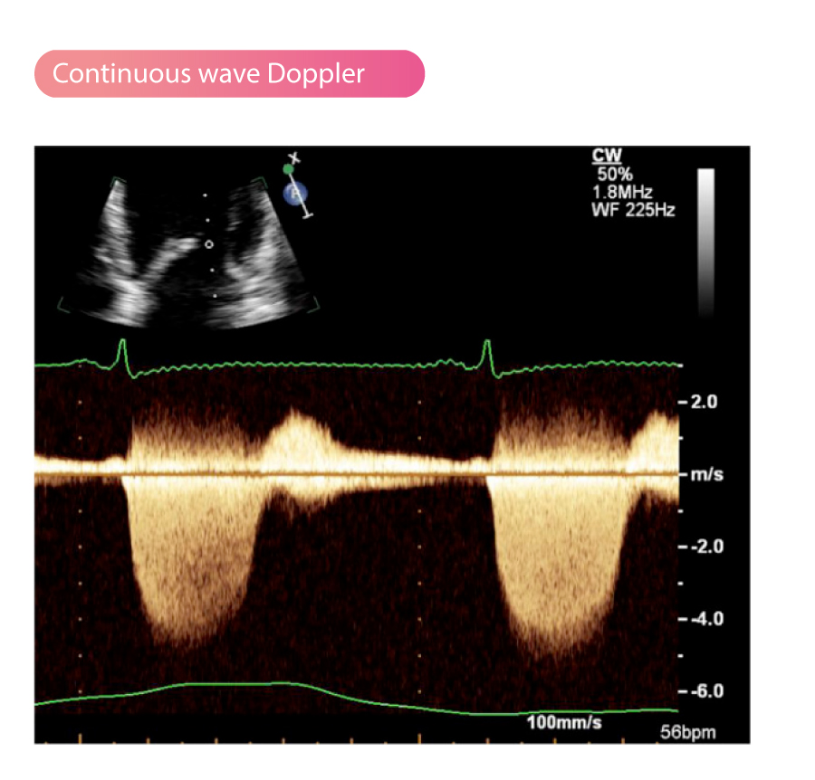
In summary, the continuous wave Doppler cannot determine the location of the maximum velocity recorded, but it allows for the recording of very high velocities along the Doppler line.

