
Catheterisation is straightforward in most patients and dangerous in a few. The male urethra is about 25 cm long with two curves and a prostate that can grip; the female urethra is only 3–5 cm and almost straight, but the meatus can be hard to find in older women. Two things separate the safe catheterisation from the harmful one: never forcing against resistance, and never catheterising urethrally when urethral injury is suspected.
Indications
- Acute urinary retention.
- Chronic urinary retention with renal impairment or a large residual volume.
- A need for accurate measurement of urine output in a critically ill patient.
- Perioperatively and postoperatively in longer procedures and with epidural anaesthesia.
- Bladder washout and continuous irrigation in macroscopic haematuria with clots (a three-way catheter).
- Instillation therapy and contrast studies of the urinary tract.
- Wound healing with a sacral pressure sore or extensive perineal wounds where urinary leakage prevents healing.
- Voiding problems in neurogenic bladder dysfunction, in the first instance with clean intermittent self-catheterisation rather than an indwelling catheter.
A catheter is not, however, indicated for incontinence as the sole reason, for convenience where the workload is heavy, or as a substitute for mobilisation and bladder training.
Contraindications
| Type | Condition |
|---|---|
| Absolute | Suspected urethral injury — blood at the meatus, a perineal or scrotal haematoma, a high-riding or impalpable prostate, a pelvic fracture, a straddle injury |
| Relative | Recent urethral or prostatic surgery — contact a urologist before attempting |
| Relative | A known urethral stricture |
| Relative | Acute prostatitis or urethritis |
| Relative | Latex allergy (choose an all-silicone catheter) |
Where urethral injury is suspected, no urethral catheter may be passed. An attempt can convert a partial rupture into a complete one. Contact the urologist or the surgeon on call for urethrography and suprapubic drainage.
Preparation and equipment
A sterile catheterisation pack, sterile gloves, chlorhexidine solution 0.2 mg/mL or an equivalent cleansing solution, sterile gauze, sterile water for the balloon, a drainage bag and a catheter valve, and lubricant containing a local anaesthetic (lidocaine gel) in a pre-filled syringe.
Catheter size is given in Charrière (Ch), where the figure corresponds to the circumference in millimetres — a Ch 16 therefore has a diameter of about 5 mm.
| Indication | Size | Comment |
|---|---|---|
| Clear urine, routine catheterisation in a woman | Ch 12–14 | A short catheter, about 20–25 cm |
| Clear urine, routine catheterisation in a man | Ch 14–16 | Standard length, about 40 cm |
| Cloudy urine, slight haematuria without clots | Ch 16 | |
| Markedly cloudy urine or sediment | Ch 18 | Prescribed by a doctor |
| Macroscopic haematuria with clots | Ch 20–24, a haematuria or three-way catheter | Large eyes, allowing irrigation |
| Prostatic enlargement, difficult passage | Ch 16–18 Tiemann | A curved, tapering tip |
A larger catheter is not easier to pass. With a difficult passage it is the shape of the tip and the technique that matter, not the calibre; choose instead the smallest size that will do the job, to reduce urethral trauma.
The balloon is filled with sterile water according to the volume printed on the catheter, usually 10 mL in adults. Do not use saline — the salt can crystallise and block the valve. The material determines how long the catheter may stay: all-silicone and latex with a hydrogel or silicone elastomer coating may remain for up to twelve weeks, latex with a PTFE coating for a shorter period.
Procedure
In men
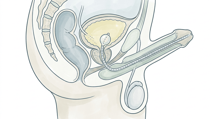
- Supine with the legs slightly apart. Create a sterile field and lay out the equipment.
- Retract the foreskin and clean the glans and meatus with the cleansing solution in circular movements from the centre outwards. Change the swab between strokes.
- Instil 10–20 mL of lidocaine gel into the urethra. Pinch the meatus closed and wait at least 3–5 minutes — the anaesthetic and lubricating effect of the gel is the single most important prerequisite for a painless catheterisation in a man.
- Hold the penis stretched upwards and forwards at about 60–90 degrees to the abdomen. This straightens the penile curve.
- Advance the catheter slowly and steadily. At resistance at the pelvic floor: lower the penis towards the legs and ask the patient to breathe calmly or to bear down gently so that the sphincter relaxes.
- Advance the catheter all the way to the bifurcation before the balloon is inflated, even if urine appears sooner. The balloon must never be inflated in the prostatic urethra.
- Inflate the balloon with the prescribed amount of sterile water. Stop immediately if the patient has pain during inflation — that indicates malposition.
- Withdraw the catheter gently until the balloon rests against the bladder neck, connect the bag and replace the foreskin over the glans.
In women
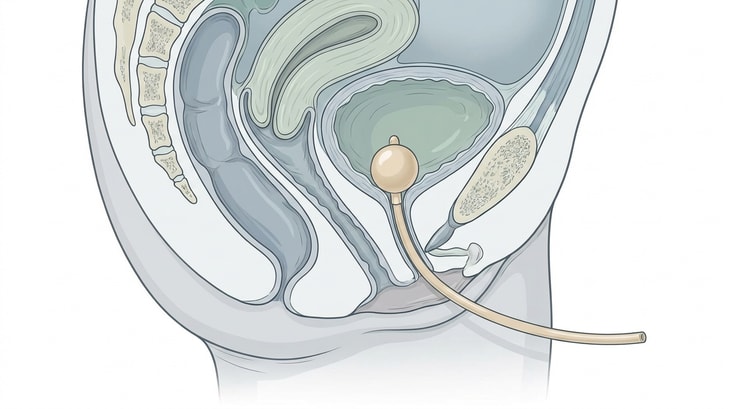
- Supine with the knees flexed and apart (the frog position), or the lateral position with the knees drawn up if the patient cannot lie on her back. Make sure the lighting is good.
- Part the labia with the non-dominant hand and maintain the grip throughout the procedure. Clean from front to back, one stroke per swab, the outer parts first and the meatus last.
- Identify the meatus — it lies between the clitoris and the vaginal opening and in older women can be retracted or hidden by atrophic mucosal folds. Draw the anterior vaginal wall gently upwards and forwards if it is hard to see.
- Instil 5–10 mL of lidocaine gel into the urethra, or lubricate the catheter generously.
- Advance the catheter until urine appears, then a further 3–5 cm so that the balloon is certainly in the bladder.
- Inflate the balloon, withdraw it against the bladder neck and connect the bag.
- A catheter in the vagina: leave it in place as a marker, take a new sterile catheter and place it in the urethra above. Only then is the misplaced one removed.
Difficult catheterisation in a man
Commonest in benign prostatic hyperplasia, where the prostatic lobes compress the urethra, and in urethral stricture after previous catheterisation or instrumentation.
- Give more gel and more time. Insufficient time for the anaesthetic is the commonest reason for an "impossible" catheterisation.
- Change to a Tiemann catheter Ch 16–18. Its tapering, upturned tip follows the curvature of the urethra over the prostatic lobe. The tip must point upwards throughout the insertion — the mark or ridge on the outer end of the catheter shows the direction of the tip.
- Have the patient bear down gently, or ask him to relax the pelvic floor, at resistance in the membranous urethra.
- Make no more than two attempts. Never force, and never use a stylet or guidewire without urological expertise — a false passage through the prostate causes bleeding, infection and later stricture.
- If the obstruction persists: contact a urologist. The options are catheterisation over a guidewire under cystoscopic control, or a suprapubic catheter, inserted percutaneously above the symphysis into a full, ultrasound-verified bladder. A suprapubic catheter is also the first choice in urethral injury and may be considered where a catheter is needed long term, since it causes fewer urethral complications.
Drainage with a large residual volume
With a very large residual volume, of the order of more than 1,000 mL, there is a tradition of draining in stages, for example 500 mL at a time with a pause, by clamping the catheter, in order to reduce the risk of haematuria ex vacuo when the vessels of the compressed mucosa are suddenly decompressed. The evidence that staged drainage really reduces the complications is weak, and practice differs between regions — several modern procedures permit complete drainage straight away.
Whichever method is used: expect a post-obstructive diuresis. The concentrating ability of the kidney is impaired and the urine output can be several litres a day over the following days, with a risk of hypovolaemia and electrolyte disturbance. Measure the hourly urine output, check the electrolytes and creatinine, and replace fluid in marked polyuria.
Complications
- Urethral injury and a false passage from forced insertion, often followed by bleeding, infection and later stricture.
- Inflation of the balloon in the urethra with urethral rupture — the most painful and damaging technical error.
- Catheter-associated urinary tract infection. The risk increases with every catheter day; bacteriuria is almost the rule after a week or so.
- Bladder spasm, bypassing alongside the catheter, and discomfort.
- Haematuria ex vacuo and post-obstructive diuresis after decompression of a large residual volume.
- Meatal necrosis from traction or from too large a catheter.
- Catheter blockage by clots or encrustation.
- Paraphimosis if the foreskin is not replaced after the procedure.
Aftercare and follow-up
- Asymptomatic bacteriuria in a catheterised patient must not be treated, and a urine culture must not be taken routinely in the absence of symptoms. Antibiotics are given for fever, systemic illness or flank pain — not for cloudy or foul-smelling urine.
- Antibiotic prophylaxis before a catheter change is not given routinely.
- A closed system: never disconnect the catheter from the bag unnecessarily, keep the bag below bladder level, and do not let it lie on the floor.
- Daily washing of the genital area with soap and water is sufficient; daily disinfectant has no demonstrated benefit.
- Review the indication daily. Catheter days that no one can any longer justify are the commonest cause of catheter-associated infection.
- Document the indication, the catheter size and material, the balloon volume, the volume of urine drained, the planned duration and the date for change or review.
- On removal after urinary retention: check that the patient can void and measure the residual volume with a bladder scan after a couple of voids.
Common pitfalls
- Catheterising despite blood at the meatus. That is a urethral injury until proven otherwise.
- Too short a time for the lidocaine gel to work. Three to five minutes, timed by the clock.
- Inflating the balloon as soon as urine appears in a man — the tip may then still be in the prostatic urethra. Advance to the bifurcation first.
- Forcing against resistance, or resorting to a larger catheter in the belief that it will push through.
- Forgetting to replace the foreskin, with paraphimosis a few hours later as the result.
- Removing a misplaced catheter from the vagina before the new one is in place — the misplaced catheter is a useful landmark.
- Saline in the balloon instead of sterile water.
- Antibiotics for cloudy urine without symptoms in a catheterised patient.
- Leaving the catheter in place without a current indication.

