
The commonest error in paracentesis is not performing it. Every patient admitted with ascites should undergo a diagnostic paracentesis, because spontaneous bacterial peritonitis often lacks abdominal pain and peritonism and instead presents as encephalopathy, renal failure or simply deterioration. The procedure itself is straightforward: ultrasound to find the fluid, an insertion site in the left lower quadrant lateral to the rectus sheath, and a cell count plus albumin on the sample.
Indications
Diagnostic paracentesis:
- Newly detected ascites — always.
- Every admission of a patient with known ascites.
- Clinical deterioration in a cirrhotic patient with ascites: fever, abdominal pain, encephalopathy, rising creatinine, gastrointestinal bleeding, leucocytosis or acidosis.
- Suspected malignancy, tuberculous peritonitis or pancreatic ascites.
Therapeutic paracentesis:
- Tense ascites with abdominal discomfort, early satiety or respiratory compromise.
- Diuretic-refractory or diuretic-intolerant ascites, as recurrent treatment.
Contraindications
There are in practice no absolute contraindications; suspected spontaneous bacterial peritonitis carries more weight than a deranged coagulation profile. Avoid or modify in:
| Situation | Management |
|---|---|
| Disseminated intravascular coagulation or clinically overt bleeding | Refrain until the condition has been corrected |
| Marked coagulopathy or thrombocytopenia | Routine plasma or platelets are not needed — the INR does not predict bleeding risk in paracentesis. Follow the local protocol |
| Ileus with distended bowel loops | Ultrasound guidance mandatory; choose another site |
| Scars, hernia or visible abdominal wall collaterals (caput medusae) | Puncture at least a few centimetres away from them |
| Full bladder | Have the patient void, or catheterise, before puncture |
| Pregnancy, previous extensive abdominal surgery | Ultrasound guidance; choose a new site if in doubt |
| Cellulitis over the insertion site | Change the puncture site |
Preparation and equipment
- Ultrasound with a curved array probe.
- Sterile preparation (chlorhexidine in alcohol 5 mg/mL), sterile gloves, fenestrated drape; full sterile attire for therapeutic drainage with an indwelling catheter.
- Lidocaine 10 mg/mL, 5–10 mL, 25 G for the skin wheal and 21 G for the deeper layers.
- Diagnostic tap: 21–22 G needle, 20 mL and 50 mL syringes.
- Therapeutic drainage: a commercial paracentesis set or a catheter over needle, 16–18 G, connected to tubing and a collection container.
- Sample tubes: an EDTA tube for the cell count, a serum tube for albumin, protein and LDH, and two blood culture bottles (aerobic and anaerobic) inoculated at the bedside.
- Albumin solution 200 mg/mL for a planned large-volume tap.
- A blood sample at the same time: serum albumin is mandatory in order to be able to calculate the SAAG.
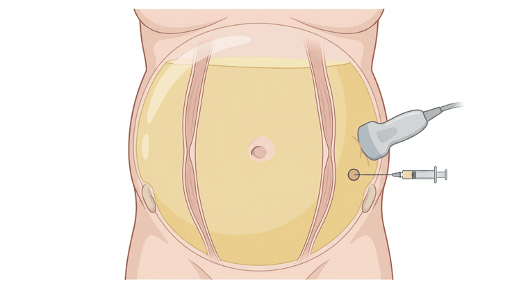
The patient lies supine, preferably with slight rotation towards the left side so that the fluid collects in the left flank. Have the patient empty the bladder first.
Procedure
Choice of insertion site
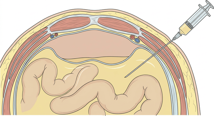
The left lower quadrant is the first choice: the abdominal wall is thinner there than on the right, and the descending colon is fixed, whereas the caecum on the right is mobile and often gas-filled. The site lies about 3–5 cm superior and medial to the anterior superior iliac spine and lateral to the lateral border of the rectus abdominis — this avoids the inferior epigastric artery, which runs on the posterior aspect of the rectus muscle along its lateral border and is the commonest source of serious bleeding after paracentesis.
The midline 2 cm below the umbilicus is an alternative in the relatively avascular linea alba, but requires an empty bladder and is rarely used in large volume ascites.
Diagnostic paracentesis
- Scan the left lower quadrant. Confirm at least 2–3 cm of free fluid depth with no bowel in the path of the needle and measure the skin-to-peritoneum distance.
- Mark the site. Sterile preparation and draping.
- Anaesthetise the skin and abdominal wall down to the peritoneum; aspirate as the needle is advanced.
- Use the Z-technique: pull the skin 2 cm to one side with the free hand, insert the needle, and release the skin as the needle is withdrawn. The tracks in skin and peritoneum are then offset and leakage afterwards becomes less common.
- Advance the needle slowly, in small steps, with continuous aspiration. A clear give in resistance followed by fluid in the syringe means the peritoneum has been passed.
- Aspirate 30–50 mL. Distribute into the tubes and inoculate the blood culture bottles directly at the bedside, 10 mL into each — this increases the bacteriological yield considerably compared with tubes sent to the laboratory.
- Withdraw the needle, compress briefly and apply a dressing.
Therapeutic (large-volume) paracentesis
- Localise and anaesthetise as above.
- Insert the catheter over the needle or by Seldinger technique and remove the stylet as soon as fluid appears. Never leave a steel needle in place — the bowel may be injured as the abdominal wall collapses.
- Connect the tubing and collection container. The fluid usually drains under the pressure of the abdomen itself; have the patient rotate slightly towards the puncture side when the flow diminishes.
- Drain until the flow of fluid ceases. There is no upper volume limit as long as albumin is given — the whole volume may be drained on one occasion.
- Monitor blood pressure and pulse during the drainage.
- Remove the catheter and apply a pressure dressing. A stoma dressing or bag may be needed if there is subsequent leakage.
Albumin replacement
When more than 5 litres are drained, albumin should be given to prevent paracentesis-induced circulatory dysfunction — a hypovolaemic, vasodilated reaction within the first 24 hours that increases the risk of hyponatraemia, hepatorenal syndrome and death.
| Volume drained | Replacement |
|---|---|
| Less than 5 litres | Albumin is generally not needed |
| More than 5 litres | 6–8 g albumin per litre drained, given during or immediately after the drainage |
With albumin solution 200 mg/mL, 100 mL contains 20 g of albumin. A 10-litre drainage therefore corresponds to about 60–80 g, that is 300–400 mL of the 200 mg/mL solution. Synthetic plasma expanders are not equivalent and are not recommended in this situation.
Interpretation
SAAG — serum-ascites albumin gradient
flowchart TD
  A[SAAG = serum albumin minus ascites albumin] --> B{SAAG 11 g/L or more?}
  B -- Yes --> C[Portal hypertension]
  C --> D{Total protein in the ascites?}
  D -- Below 25 g/L --> E[Liver cirrhosis, acute liver failure, massive liver metastases]
  D -- 25 g/L or more --> F[Heart failure, constrictive pericarditis, Budd-Chiari]
  B -- No --> G[No portal hypertension]
  G --> H[Peritoneal carcinomatosis, tuberculous peritonitis, pancreatic ascites, nephrotic syndrome, serositis]
The SAAG is calculated on samples taken at the same time. The gradient distinguishes portal hypertension from other causes with high accuracy and is not affected by diuretic treatment. A low albumin in the ascites is not the same as a low SAAG — it is the difference from serum that counts.
Spontaneous bacterial peritonitis
The diagnosis rests on the polymorphonuclear cell count in the ascites — 250 × 10⁶/L or more — irrespective of the culture result. Antibiotics are started on the cell count; about half of cases are culture negative.
- With blood-stained ascites: subtract 1 polymorphonuclear cell per 250 erythrocytes before applying the threshold.
- More than one bacterial species in the culture, a very high cell count, glucose below 2.8 mmol/L, protein above 10 g/L or LDH above the serum level suggest secondary bacterial peritonitis — a surgical diagnosis that requires computed tomography, not antibiotics alone.
- The choice of antibiotic and the doses follow the local protocol; in Sweden intravenous cefotaxime is usually the first-line agent.
- Albumin in addition to antibiotics reduces the risk of hepatorenal syndrome and death in spontaneous bacterial peritonitis: 1.5 g/kg on the first day and 1 g/kg on day three.
Other analyses
| Analysis | Comment |
|---|---|
| Cell count and differential (EDTA tube) | The basic analysis; sent without delay |
| Albumin (together with a simultaneous serum albumin) | For the SAAG |
| Total protein | Distinguishes cardiac from cirrhotic ascites; a value below 15 g/L identifies a high risk of spontaneous bacterial peritonitis |
| Culture in blood culture bottles | Inoculated at the bedside |
| Cytology | When malignancy is suspected; requires a large volume and repeated samples |
| Amylase | Markedly raised in pancreatic ascites |
| Triglycerides | With milky fluid (chylous ascites) |
| Mycobacterial culture, PCR, adenosine deaminase | When tuberculous peritonitis is suspected; low sensitivity, laparoscopy with biopsy gives a higher yield |
Complications
| Complication | Comment |
|---|---|
| Persistent fluid leakage from the insertion site | The commonest; countered by the Z-technique |
| Abdominal wall haematoma | Usually mild |
| Haemoperitoneum after injury to the inferior epigastric artery | The most serious; avoided by an insertion site lateral to the rectus sheath |
| Bowel perforation | Rare; increased risk in ileus and adhesions — ultrasound before puncture |
| Infection of the abdominal wall or iatrogenic peritonitis | Sterile technique |
| Paracentesis-induced circulatory dysfunction | After large-volume drainage without albumin |
| Hyponatraemia and renal failure | Follow electrolytes and creatinine after large-volume taps |
Aftercare and follow-up
- Check blood pressure and pulse after the tap; follow sodium and creatinine the day after a large-volume drainage.
- With subsequent leakage: a pressure dressing, a stoma bag over the insertion site and lateral positioning with the puncture site uppermost. A single suture may be needed for persistent leakage.
- Treat the underlying disease: salt restriction to about 5–6 g of sodium chloride per day and diuretics (spironolactone with the addition of furosemide) are the mainstay in cirrhotic ascites. Fluid restriction is needed only in hyponatraemia.
- Stop NSAIDs, and be restrictive with high-dose beta blockers and with ACE inhibitors and ARBs in refractory ascites and low blood pressure.
- Consider antibiotic prophylaxis after an episode of spontaneous bacterial peritonitis and when the protein content of the ascites is very low, according to the local protocol.
- When large-volume drainage is repeatedly required: refer for consideration of TIPS and assessment for liver transplantation.
Common pitfalls
- Not tapping at all. Omitting the diagnostic paracentesis on admission is the single commonest and most serious failing.
- An insertion site through the rectus sheath instead of lateral to it — directly over the inferior epigastric artery.
- Puncture in the right lower quadrant where the gas-filled caecum is often in the way.
- Puncture through scars or visible collaterals.
- Forgetting the simultaneous serum albumin — the SAAG can then not be calculated.
- Culture sent in a tube instead of inoculated into blood culture bottles at the bedside.
- Waiting for the culture result before starting antibiotics when the polymorphonuclear cell count is above 250 × 10⁶/L.
- Large-volume drainage without albumin.
- Routine plasma or platelets before paracentesis in a cirrhotic patient without active bleeding.
- Missing secondary peritonitis and treating a surgical abdomen with antibiotics alone.

