
A monoarthritis is septic until proven otherwise, and the only thing that proves otherwise is the synovial fluid. The aspiration is technically simple — what determines the outcome is that it is done early and sterilely and that the samples are sent correctly. A corticosteroid must never be injected into a joint in which infection has not been excluded.
Indications
- Acute monoarthritis in which septic arthritis must be distinguished from crystal arthritis — the most important indication and an emergency procedure.
- An unclear arthritis, as part of the diagnostic work-up.
- Haemarthrosis after trauma.
- A symptomatic effusion causing pain or restricted movement (therapeutic aspiration).
- Intra-articular corticosteroid injection in verified inflammatory or osteoarthritis-related joint disease.
Contraindications
- Infection of the skin or soft tissues over the puncture site — use another approach or refrain.
- Bacteraemia without suspicion of joint infection (relative; aspiration can seed a sterile joint).
- Marked coagulopathy or an anticoagulant at a therapeutic dose (relative; weigh the risk against the diagnostic benefit — where sepsis is suspected, the diagnosis always weighs more heavily).
- A joint prosthesis: the aspiration must be performed by an orthopaedic surgeon under strictly sterile conditions.
For corticosteroid injection there is an additional absolute contraindication: the slightest suspicion of septic arthritis. If the picture is unclear — aspirate, send a culture, await the result and inject later.
Preparation and equipment
- Chlorhexidine in alcohol 5 mg/mL: clean with 3–4 swabs in wide circles and let it dry for a couple of minutes before the needle is inserted.
- Sterile gloves, sterile gauze, and a fenestrated drape for injection.
- A needle of 0.8–1.2 × 50 mm for large joints (knee, shoulder), 0.5–0.6 × 25 mm for small joints (wrist, finger joints).
- A 20–50 mL syringe for a knee with a large effusion, 5–10 mL for smaller joints.
- Local anaesthesia of the skin and subcutis where needed, lidocaine 10 mg/mL.
- Sample tubes: an EDTA tube for the cell count and crystals, a sterile tube and blood culture bottles (aerobic and anaerobic) for culture, and a fluoride tube for glucose and lactate.
- For injection: the corticosteroid drawn up in its own syringe, so that the same needle can stay in place when the syringe is changed after aspiration.
Procedure
- Examine the joint, locate the effusion and mark the puncture site with a pen or an indentation from a fingernail before cleaning the skin.
- Clean and let it dry. Do not palpate the puncture site again with an unsterile hand.
- Insert the needle in one movement, aspirating gently as it advances. Fluid in the syringe confirms the position.
- Empty the joint as completely as possible — compress the bursa with your hand to press the remaining fluid down.
- If a corticosteroid is to be given: hold the needle still, change the syringe and inject. The injection should go in easily. Resistance means the tip is in tissue, not in the joint cavity.
- Withdraw the needle, apply compression and a dressing.
The knee
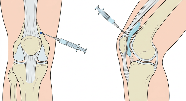
The patient lies supine with the leg extended and the quadriceps relaxed. The superolateral approach has the highest accuracy for palpation-guided aspiration. Press the fluid from the medial towards the lateral side with your free hand. The needle is inserted directed medially and slightly caudally, parallel to the posterior surface of the patella.
The shoulder
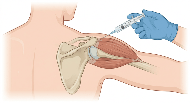
The patient sits with the arm hanging relaxed and slightly internally rotated. The posterior approach is preferable: it passes through muscle without major vessels or nerves, and the landmark — the posterior corner of the acromion — is easy to palpate even in a large patient. The anterior approach, just lateral to the coracoid process, is an alternative but lies closer to the brachial plexus and the vessels.
The elbow
The elbow is flexed to 90 degrees with the palm facing down. Palpate the triangle between the lateral epicondyle, the tip of the olecranon and the radial head — the joint is most superficial there. The needle is inserted perpendicularly in the centre of the triangle, directed medially. Ask the patient to pronate and supinate slightly to identify the radial head.
The wrist
A dorsal approach with the wrist slightly flexed over a roll. The puncture site lies just distal to the distal end of the radius, ulnar to Lister's tubercle, in line with the third metacarpal. Use a fine needle and a shallow depth — the radiocarpal joint is narrow and the yield is often only a few drops. Avoid the extensor tendons.
The ankle
An anteromedial approach with the foot in slight plantar flexion. The puncture site lies in the groove between the medial malleolus and the tibialis anterior tendon, above the dome of the talus. The needle is directed laterally and slightly dorsally. Avoid the dorsalis pedis artery and vein and the deep peroneal nerve, which lie lateral to the tendon.
Handling and interpretation of the sample
Always assess the fluid macroscopically first: volume, colour, clarity and viscosity. Cloudy, thin fluid suggests inflammation; blood with fat droplets suggests an intra-articular fracture.
| Tube | Analysis | Volume |
|---|---|---|
| EDTA tube, well mixed | Cell count and differential, crystals under polarised light | 2–5 mL |
| Sterile tube and blood culture bottles (aerobic and anaerobic) | Culture and direct microscopy with Gram staining | 2–5 mL |
| Fluoride tube | Glucose and lactate, with a simultaneous blood sample for the ratio | A few mL |
| Type of synovial fluid | White cell count (×10⁹/L) | Granulocytes | Typical of |
|---|---|---|---|
| Normal | < 0.2 | < 25 % | — |
| Non-inflammatory | < 2 | < 25 % | Osteoarthritis, trauma |
| Inflammatory | 2–50 | 25–70 % | Rheumatoid arthritis, reactive arthritis |
| Crystal arthritis | 2–80 | 50–70 % | Gout, calcium pyrophosphate arthritis |
| Septic | (50–)70–200 | > 70 % | Bacterial arthritis |
flowchart TD
  A[Acute monoarthritis] --> B[Joint aspiration, cell count, crystals, culture]
  B --> C{White cell count in the synovial fluid}
  C -- Below 2 --> D[Non-inflammatory: osteoarthritis or trauma]
  C -- 2 to 50 --> E{Crystals?}
  E -- Yes --> F[Crystal arthritis likely. Culture anyway]
  E -- No --> G[Inflammatory arthritis. Follow clinically and await the culture]
  C -- Above 50, granulocytes above 70 per cent --> H[Treat as septic arthritis]
  H --> I[Culture, drainage and antibiotics. No corticosteroid injection]
  F --> J{Fever, a high CRP or a patient who is systemically unwell?}
  J -- Yes --> H
  J -- No --> K[Treat the crystal arthritis, re-aspirate if the patient deteriorates]
A finding of crystals does not exclude infection. Gout and septic arthritis can be present in the same joint at the same time, and a high cell count and a raised lactate are also seen in gout and in a flare of rheumatoid arthritis. It is the culture and the direct microscopy that decide. Conversely, a negative direct microscopy does not exclude septic arthritis — where there is clinical suspicion, the patient is treated while the culture result is awaited.
Drugs for injection
- Methylprednisolone acetate (Depo-Medrol) 40 mg/mL: 20–80 mg (0.5–2 mL) into large joints according to the Swedish medicines compendium (FASS), with a lower dose into smaller joints.
- Triamcinolone hexacetonide (Lederspan) is used in large joints as an alternative; the dose is according to FASS and local practice.
- Mixing with a local anaesthetic is practised and varies between units.
- The effect appears after a day or so and typically lasts 3–4 weeks. Repeated injections into the same joint should be limited; several injections a year into a weight-bearing joint increase the risk of cartilage and tendon damage.
Complications
- Iatrogenic septic arthritis — rare but serious; the risk is linked to inadequate sterile technique.
- Haemarthrosis, particularly with anticoagulant treatment.
- A dry tap with no yield.
- Cartilage damage from the needle tip.
- After corticosteroid injection: a post-injection flare with increased pain for 1–2 days, subcutaneous atrophy and hypopigmentation when the injection is superficial, tendon rupture if injected into a tendon, and a transient rise in blood glucose in patients with diabetes.
Aftercare and follow-up
A pressure dressing and rest for the first 24 hours after aspiration of a large joint. After corticosteroid injection, relative rest of the joint for 1–2 days is recommended. Tell the patient to seek urgent help if the pain, redness or swelling increases or if fever develops after the procedure — that is how an iatrogenic infection presents.
In septic arthritis the joint must be drained repeatedly or surgically, and the patient managed together with an orthopaedic surgeon and an infectious diseases physician. In diabetes: check the blood glucose extra carefully for the next few days after a corticosteroid injection.
Common pitfalls
- A corticosteroid in a joint that turns out to be septic. At the slightest uncertainty: aspirate, culture and wait.
- Postponing the aspiration in acute monoarthritis while awaiting radiography or laboratory results.
- A culture that is not taken when the question at issue is septic arthritis after all.
- Trusting the finding of crystals and dismissing infection.
- Too fine a needle in the knee — the fluid is often viscous and the needle blocks.
- Synovial fluid in the wrong tube. The cell count cannot be performed on a clotted sample without EDTA.
- Aspiration through infected skin, which can introduce bacteria into a sterile joint.

