
Aspirate and biopsy answer different questions and do not replace one another: the aspirate shows cellular morphology and provides material for flow cytometry and genetics, while the biopsy shows marrow architecture and cellularity. What usually ruins the examination is not the technique but the handling of the specimen — an aspirate that clots before the smears are made is unusable, no matter how well the puncture went.
Indications
- Unexplained cytopenia in one or more cell lines.
- Suspected acute leukaemia, myelodysplastic syndrome or myeloproliferative neoplasm.
- Work-up and staging of lymphoma and myeloma.
- Suspected bone marrow metastasis.
- Assessment of treatment response and measurement of residual disease (MRD).
- Fever of unknown origin or suspected granulomatous disease with marrow involvement.
- Work-up of unexplained iron, B12 or folate deficiency when blood tests are insufficient.
Contraindications
- Severe coagulopathy or ongoing anticoagulation — correct this first. Thrombocytopenia, by contrast, is not a contraindication; the specimen may be taken even at very low platelet counts, followed by compression.
- Infection in the skin overlying the puncture site.
- Previous pelvic irradiation on the intended side (yields fibrotic, unrepresentative marrow — choose the other side).
- A patient who cannot lie still — plan for sedation rather than abandoning the procedure.
Choice of site
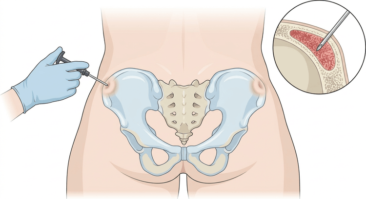
The posterior superior iliac spine is the standard site for both aspiration and biopsy in adults. Here the posterior part of the iliac crest is thick, red marrow is abundant, and the distance to vessels and abdominal organs is large.
- The sternum may be used for aspiration when the iliac crest is inaccessible, but biopsy must never be taken from the sternum — the needle may perforate into the mediastinum.
- The anterior superior iliac spine is a second-line alternative in marked obesity or when the supine position is necessary.
- After previous punctures: change side, and avoid irradiated fields.
Preparation and equipment
- Informed consent, current full blood count and coagulation screen, medication list.
- Chlorhexidine in alcohol, sterile gloves, fenestrated drape, sterile gauze.
- Local anaesthetic: lidocaine 10 mg/mL, usually 5–10 mL, with a longer needle so that the periosteum can be infiltrated generously — it is the periosteum that hurts, not the skin.
- Aspiration needle (iliac crest or sternal needle with stylet) and a Jamshidi-type biopsy needle.
- Syringes: a small 5–10 mL syringe for the first aspirate, larger syringes for the tube specimens.
- Glass slides, at least 6–10, plus spreader slides — laid out and labelled before the puncture begins.
- Specimen tubes: EDTA and heparin tubes according to local instructions, plus a formalin container for the biopsy.
- No. 11 scalpel for the skin incision, sterile dressings, pressure dressing.
Procedure
- Place the patient in the lateral position with the knees drawn up, or prone with a pillow under the pelvis. The lateral position is usually the most comfortable and gives good access.
- Palpate the iliac crest posteriorly until you feel the rounded prominence that is the posterior superior iliac spine. Mark it.
- Clean the skin and apply the drape. Anaesthetise skin, subcutaneous tissue and then the periosteum at several points around the puncture site, and wait a few minutes.
- Make a small skin incision with the scalpel.
- Aspiration: advance the needle with the stylet in place perpendicular to the bone surface, angled slightly anteriorly and medially. Rotate under steady pressure until the resistance suddenly gives way — the cortex has then been passed. Remove the stylet and aspirate.
- Make the smears immediately at the bedside from the first aspirate, which should be small — a few tenths of a millilitre, at most about 0.5 mL. A larger volume draws in peripheral blood and dilutes the specimen. Marrow clots within seconds; the smears must be made before that.
- Then aspirate further volume into the tubes. When the question is MRD, the reverse order often applies — the first portion is reserved for flow cytometry. Follow local instructions.
- Biopsy: take the biopsy through the same skin incision but in a slightly different direction, or a few millimetres away from the aspiration hole, so that it is not taken from an area already admixed with blood. Advance the Jamshidi needle with the stylet through the cortex, remove the stylet and advance the needle a further 2–3 cm with rotating pressure.
- Rotate the needle a couple of turns in both directions to break off the core, withdraw it while rotating, and expel the specimen backwards with the probe — never through the tip, which deforms the specimen.
- Inspect the biopsy: it should be at least 15–20 mm long and of bony consistency. A short, soft or fragmented core is often uninterpretable — repeat it.
- Gauze, firm pressure for at least 5–10 minutes, longer in thrombocytopenia, then a pressure dressing.
Specimen allocation
flowchart TD
  A[Puncture at the posterior superior iliac spine] --> B[First aspirate, at most 0.5 mL]
  B --> C[Smears immediately at the bedside, 6 to 10 slides]
  C --> D[Morphology and iron staining]
  A --> E[Further aspirate into tubes]
  E --> F[Flow cytometry: 2 to 3 mL in heparin or EDTA tube]
  E --> G[Cytogenetics and FISH per local instructions]
  E --> H[Molecular genetics and PCR]
  A --> I[Biopsy with Jamshidi needle, at least 15 to 20 mm]
  I --> J[Formalin to pathology]
J --> K[Cellularity, fibrosis, architecture and immunohistochemistry]| Specimen | Answers | Handling |
|---|---|---|
| Aspirate smears | Cellular morphology, degree of maturation, blast percentage, iron stores | Must be made within seconds, air-dried, no fixation beforehand |
| Aspirate in tubes | Immunophenotype by flow cytometry, chromosome analysis, molecular diagnostics | 2–3 mL per assay, to the laboratory without delay |
| Biopsy in formalin | Cellularity, marrow architecture, fibrosis, focal infiltrates, immunohistochemistry | The only specimen that yields an answer in a dry tap |
A dry tap — no aspirate can be obtained — is itself a finding and suggests myelofibrosis, hairy cell leukaemia or a heavily infiltrated marrow. In a dry tap the biopsy is mandatory, and touch preparations of the biopsy core against glass slides often provide morphological information that substitutes for the smears.
Analgesia and sedation
The procedure hurts at two moments: when the periosteum is anaesthetised and when the aspirate is drawn. The aspiration pain is brief, severe and cannot be anaesthetised away — warn the patient immediately before you draw.
- Baseline: good local anaesthesia with generous periosteal infiltration, plus paracetamol and possibly an opioid beforehand.
- Nitrous oxide (Entonox) is well established in Swedish practice, rapidly reversible and requires no monitoring beyond the usual.
- In marked anxiety, previous traumatic experience or a planned biopsy: sedation with a benzodiazepine and an opioid according to local protocol, with monitoring of oxygen saturation and respiration and reversal agents available.
- Children undergo the examination under general anaesthesia or deep sedation.
Complications
Serious complications are rare. The most common are:
- Pain and tenderness at the puncture site for a few days.
- Bleeding and haematoma — the most common serious complication, associated with thrombocytopenia, coagulopathy and myeloproliferative disease.
- Infection, very uncommon.
- Retroperitoneal bleeding or vascular injury from a misdirected or excessively advanced needle.
- Needle fracture within the bone.
- Injury to abdominal organs or great vessels when puncture is performed outside the anatomical landmarks.
- With sternal puncture: perforation with cardiac tamponade or vascular injury — the reason biopsy is never taken from that site.
Aftercare and follow-up
The pressure dressing stays on for a few hours, longer in thrombocytopenia. It is helpful for the patient to lie supine against the dressing for 15–30 minutes afterwards for compression. The dressing can be removed and the area showered after about 24 hours.
Tell the patient to contact medical services in case of increasing pain, enlarging swelling, bleeding through the dressing or fever. Paracetamol is usually enough for the after-pain. After sedation the patient must be monitored until fully recovered and must not drive that day.
Document the puncture site, the side, the number of smears, the length of the biopsy and which tubes were sent — this helps when the specimen has to be repeated or compared at follow-up.
Common pitfalls
- Smears made too late. The aspirate clots within seconds; the slides must be ready and waiting.
- Too large a first aspirate, which is diluted with peripheral blood and makes the morphology misleading.
- Inadequately anaesthetised periosteum — the most common reason the procedure is experienced as unbearable.
- A biopsy that is too short or crushed. Below 15 mm it is often uninterpretable; expulsion through the tip deforms the core.
- Biopsy taken through the same channel as the aspirate, which yields a blood-admixed specimen that is difficult to interpret.
- No biopsy in a dry tap — leaving no material at all to assess.
- Biopsy from the sternum. Never.
- Specimen tubes that do not match the laboratory's instructions — flow cytometry and cytogenetics have different requirements and different transport times.

