
The biopsy method determines what the pathologist can report. A shave biopsy of a pigmented lesion that turns out to be a melanoma can make the Breslow thickness unmeasurable and staging therefore impossible; a superficial punch in a case of bullous dermatosis misses the subepidermal split. Choose the technique according to the diagnostic question, not according to what is quickest.
Indications
- Suspected malignancy: basal cell carcinoma, squamous cell carcinoma, melanoma.
- An unclear inflammatory dermatosis in which the histology governs treatment.
- Suspected bullous dermatosis (which also requires a biopsy for direct immunofluorescence).
- Suspected cutaneous vasculitis, lymphoma or granulomatous disease.
- A treatment-resistant rash without a definite diagnosis.
Choice of technique
flowchart TD
  A[Lesion to be biopsied] --> B{Pigmented lesion with suspected melanoma?}
  B -- Yes --> C[Excision with a narrow margin down into the subcutis]
  B -- No --> D{Is deep dermis or subcutis needed for the diagnosis?}
  D -- Yes, panniculitis or a deep process --> E[Excision or a deep punch down into the fat]
  D -- No --> F{Inflammatory dermatosis or vasculitis?}
  F -- Yes --> G[4 mm punch from the active edge]
  G --> H{Bullous dermatosis or vasculitis?}
  H -- Yes --> I[An additional biopsy for immunofluorescence in Michel's medium]
  H -- No --> J[Formalin is sufficient]
  F -- No --> K{Superficial exophytic lesion without suspicion of malignancy?}
  K -- Yes --> L[Shave]
K -- No --> M[3-4 mm punch]| Diagnostic question | Method | Comment |
|---|---|---|
| Pigmented lesion, suspected melanoma | Excision with a 1–3 mm margin, down into the subcutis | Never a shave — the depth must be measurable |
| Basal cell carcinoma, squamous cell carcinoma | 3–4 mm punch or shave | Diagnostic biopsy before definitive surgery |
| Inflammatory dermatosis | 4 mm punch from the active edge, not from the centre | Include epidermis and dermis |
| Bullous dermatosis | Two biopsies: one from a fresh blister (routine), one from perilesional skin (immunofluorescence) | The immunofluorescence biopsy goes into Michel's medium, not formalin |
| Suspected vasculitis | 4 mm punch from a lesion less than 48 hours old | Also take a biopsy for immunofluorescence |
| Panniculitis | Excision or a deep punch down into the fat | A 4 mm punch is rarely sufficient |
| Superficial, exophytic lesion (fibroma, seborrhoeic keratosis) | Shave | Simple, with a good cosmetic result |
| Suspected cutaneous lymphoma | Several 4–6 mm punches | Broad sampling increases the yield |
Contraindications
There are no absolute contraindications. Take care in:
- Anticoagulant therapy — this rarely needs to be stopped for small biopsies, but have diathermy or aluminium chloride available.
- Infection in the area to be biopsied.
- Hypersensitivity to local anaesthetics.
- Areas at high risk of unsightly or hypertrophic scars: the sternum, the shoulder, the jawline, the ears.
- Anatomically hazardous areas: the branches of the facial nerve at the temple, the accessory nerve in the posterior triangle of the neck, and the superficial nerves of the forearm and the dorsum of the hand.
Preparation and equipment
- Chlorhexidine in alcohol or an iodine solution.
- Lidocaine 10 mg/mL with adrenaline 5 µg/mL (adrenaline gives a bloodless field and a longer duration; it is safe even on the fingers, toes, nose and ears), a 27 G needle, a 2–5 mL syringe.
- Punches of 2, 3, 4 and 6 mm.
- A size 15 scalpel, a double-edged razor blade or a flexible shave blade.
- Artery forceps, toothed surgical forceps, scissors, a needle holder.
- Sutures: 5-0 or 6-0 non-absorbable for the face, 4-0 for the trunk and limbs.
- Haemostasis: aluminium chloride solution 20–35 %, ferric subsulfate, electrocoagulation.
- A pot of 4 % formalin, and for immunofluorescence Michel's medium or saline according to local procedure.
Photograph and draw in the site before the local anaesthetic is given — the anaesthetic can make a subtle lesion hard to find again, and it is too late to wonder where the biopsy was taken when the report arrives.
Procedure
Punch biopsy
- Mark the biopsy site. In an inflammatory dermatosis: the active edge, not the burnt-out centre.
- Clean the skin and infiltrate the anaesthetic intradermally and subcutaneously until a weal forms.
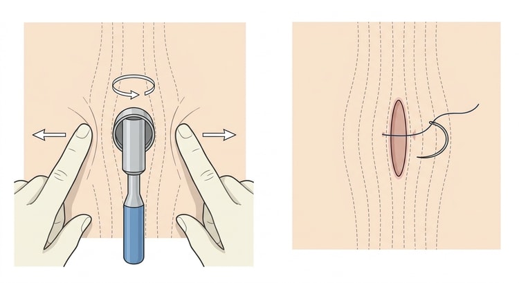
- Stretch the skin perpendicular to the skin tension lines with the thumb and index finger — the biopsy defect then becomes oval when the skin is released, which allows neater closure.
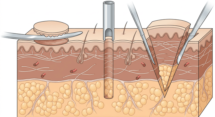
- Place the punch perpendicular to the skin and rotate under even pressure until the resistance gives way — the dermis has then been passed and you are in the subcutis.
- Lift the specimen gently with a needle tip or fine forceps applied to its edge, not by gripping across the whole biopsy, and cut the base with scissors.
- Achieve haemostasis, and place one or two sutures for 4 mm or larger. Biopsies under 3 mm can be left to heal by secondary intention.
Shave biopsy
- Infiltrate the anaesthetic so that the lesion is lifted on a cushion of anaesthetic.
- Pass the blade parallel to the skin surface in a smooth sawing movement through the base of the lesion. Bend the flexible blade to control the depth.
- Achieve haemostasis with aluminium chloride or electrocoagulation. No suture.
- The wound heals with a pale, slightly depressed patch.
Excisional biopsy
- Draw an ellipse with a length-to-width ratio of about 3:1, orientated along the skin tension lines, with the planned margin drawn in before the anaesthetic is given.
- Incise perpendicularly through the whole dermis down into the subcutis.
- Lift the specimen at one corner and dissect it free in an even plane.
- Undermine if necessary, achieve haemostasis, and close in two layers: an absorbable subcutaneous suture and a skin suture.
- Mark the orientation of the specimen with a suture and describe the marking on the request form if margin assessment is relevant.
Complications
- Bleeding and haematoma.
- Infection (low risk; prophylactic antibiotics are not needed routinely).
- Scarring, a hypertrophic scar or keloid.
- Wound dehiscence, particularly over joints.
- Pigmentary change, above all in patients with darker skin.
- Nerve injury with loss of sensation.
- An inadequate or wrongly taken specimen — the commonest complication and the one with the greatest consequences.
Aftercare and follow-up
A pressure dressing for the first 24 hours. A dry dressing, and daily cleansing with soap and water after 24 hours. Suture removal: face 5–7 days, neck 7 days, trunk 10–14 days, limbs 10–14 days, over joints 14 days.
The request form must state the exact site (preferably with a photograph or a body map), the appearance and size of the lesion, its duration and course, previous treatment, the diagnostic question, and which technique was used. A pathologist who does not know what you suspect will answer the wrong question.
Have a system that captures the report. The patient who is biopsied for suspected melanoma and then falls through the net is the single greatest risk in the whole procedure.
Common pitfalls
- A shave biopsy of a pigmented lesion — this makes the depth of invasion impossible to measure.
- A biopsy from the centre of the lesion in an inflammatory dermatosis; the edge is where the disease is active.
- Too superficial a punch that does not reach the subcutis — inadequate material for panniculitis and deep dermatoses.
- Crush injury to the specimen from forceps produces artefacts that can make assessment impossible.
- A forgotten immunofluorescence biopsy in bullous dermatosis and vasculitis — it cannot be taken retrospectively from the formalin pot.
- No documentation of the site. If the report is positive you must be able to find your way back exactly.
- No system for following up the report.

