
The result of a repair is determined before the first needle goes in. A wound that has not been irrigated clean, has not been inspected to its depth, and has not been assessed for tendon and nerve injury will heal badly however neatly the suture is placed. The commonest cause of wound infection is retained contamination, not poor suture technique — and the commonest cause of permanent loss of function is a tendon injury that was sutured over without being found.
Indications
- A traumatic wound that gapes and cannot be expected to heal with good function or cosmesis without apposition.
- A wound that bleeds because the edges are held apart.
- A wound through the full thickness of the dermis, particularly over joints and in cosmetically sensitive areas.
Superficial wounds with well apposed edges, abrasions and small wounds under about 5 mm often do better with adhesive strips, tissue adhesive or a dressing alone.
Contraindications
Primary closure must be avoided or considered carefully in:
- Bite wounds — particularly cat and human bites, and all bites on the hand. These are as a rule left open. Facial bites can be sutured after careful debridement because of the cosmesis, often with antibiotic prophylaxis.
- Deep puncture wounds, where the base cannot be inspected or irrigated clean.
- Wounds with retained foreign material or devitalised tissue that cannot be removed.
- A clinically infected wound.
- Heavily contaminated wounds where a long time has passed since the injury (see below).
Be particularly cautious with impaired healing: peripheral vascular disease, diabetes with neuropathy, immunosuppression, and high-dose corticosteroids.
The time limit and the choice of closure strategy
The classical limit of 6–8 hours for primary closure rests on weak evidence. More recent data suggest that clean, well cleaned wounds can be closed later than that, and on the face and scalp — where the blood supply is good and a scar is stigmatising — up to about 24 hours is accepted in Swedish practice. The limit is not the clock but the degree of contamination, the depth and site of the wound, and how well the wound can be cleaned. Practice differs between units; follow the local care programme where one exists.
flowchart TD
  A[A traumatic wound] --> B[Copious irrigation and inspection of the base of the wound]
  B --> C{Retained dirt, dead tissue or a foreign body?}
  C -- Yes --> D[Debride. If that is not possible: leave it open]
  C -- No --> E{Signs of infection, a bite wound or a deep puncture wound?}
  E -- Yes --> F[Leave it open]
  E -- No --> G{A clean wound, an acceptable time since the injury?}
  G -- Yes --> H[Primary closure]
  G -- No --> I[Delayed primary closure after 3–5 days]
  F --> J[Healing by secondary intention, or delayed primary closure]
  D --> J
Preparation and equipment
Assess and document before the local anaesthetic: distal sensation, motor function, capillary refill and pulse, and active movement of every tendon crossing the wound. A tendon injury can be painless and gives normal passive movement — test actively against resistance, tendon by tendon. With a glass injury: consider radiography, since glass is usually visible on plain films.
- Irrigation fluid: a copious volume of drinking-quality tap water or sodium chloride 9 mg/mL. The volume matters more than the fluid; irrigate under pressure through a 20 mL syringe and a large-bore needle.
- Chlorhexidine in alcohol to the skin around the wound — not into the wound.
- Sterile gloves, a fenestrated drape, toothed forceps, a needle holder, scissors, artery forceps, a size 15 scalpel.
- Local anaesthetic, suture material, scissors, dressings.
Local anaesthesia
| Agent | Onset | Duration | Comment |
|---|---|---|---|
| Lidocaine 10 mg/mL | Rapid | 1–2 hours | The standard choice |
| Lidocaine 10 mg/mL with adrenaline 5 µg/mL | Rapid | 2–6 hours | A bloodless field, less bleeding |
| Mepivacaine 10 mg/mL | Rapid | Somewhat longer than lidocaine | A common alternative |
| Bupivacaine 2.5 mg/mL | Slow | Long | When prolonged analgesia is wanted |
The maximum dose of lidocaine is given in the Swedish medicines compendium (FASS) and in Swedish anaesthetic reviews as about 3–4 mg/kg without adrenaline and 7 mg/kg with adrenaline. Adrenaline gives a bloodless field and a longer duration. The old rule that adrenaline is forbidden on fingers, toes, nose, ears and penis has been abandoned in the modern literature for commercial preparations at normal concentrations; be cautious nonetheless in known peripheral vascular disease and follow local practice.
Inject through the wound edge, not through intact skin — it hurts considerably less. Inject slowly. For wounds on a finger, use a digital nerve block.
Procedure
- Anaesthetise, clean the skin around the wound and apply the drape.
- Irrigate copiously. As a guide: at least 50–100 mL per centimetre of wound length, and more for dirty wounds.
- Inspect the base of the wound in a bloodless field with good lighting. Look for a foreign body, joint capsule, tendon sheath, and bone surface.
- Debride: excise clearly devitalised tissue and trim ragged wound edges sparingly so that they become straight and perpendicular to the skin.
- Close the deep layer where needed with an absorbable subcutaneous suture that takes tension off the skin suture and obliterates dead space. Avoid this in contaminated wounds — every knot is a foreign body.
- Appose the skin. Place the first suture in the middle of the wound and then halve the remaining distances.
- The needle is inserted perpendicular to the skin, curves down into the dermis and takes more tissue at depth than at the surface — this everts the wound edges, which is the aim. The entry point is 3–5 mm from the edge on the trunk and limbs, and 2–3 mm on the face.
- Tie so that the edges lie edge to edge without blanching. A suture that is tight is a suture that causes necrosis and stitch marks. The knot is placed to one side of the wound line.
- A dry dressing. For a wound over a joint: immobilise.
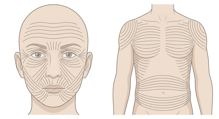
Suture techniques
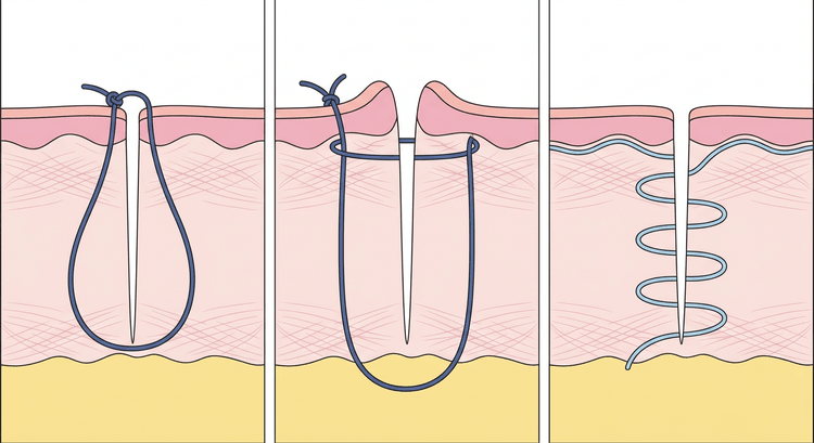
- The simple interrupted suture — the first choice for most traumatic wounds. Easy to place, and easy to remove one at a time if the wound becomes infected.
- The vertical mattress suture — for wounds under tension or where the edges invert. It gives strong eversion but more stitch marks; remove it early.
- The horizontal mattress suture — haemostasis in the scalp, and taking tension off fragile skin.
- The subcutaneous absorbable suture — takes tension off the skin suture and obliterates dead space in deep wounds.
- The continuous subcuticular suture — the best cosmesis in clean, tension-free wounds. Unsuitable in contaminated wounds.
Choice of suture material
| Site | Gauge | Suture removal |
|---|---|---|
| Face, eyelid | 6-0 | 5 days |
| Neck | 5-0 | 5–7 days |
| Scalp | 3-0 or 4-0 | 7–10 days |
| Trunk | 3-0 or 4-0 | 10–14 days |
| Upper limb, hand | 4-0 or 5-0 | 10–14 days |
| Lower limb | 3-0 or 4-0 | 14 days |
| Sole of the foot, over a joint | 3-0 | 14 days or longer |
Use a monofilament non-absorbable thread (polypropylene, polyamide) for skin — braided thread harbours bacteria. Absorbable thread in the deep layers. In children and in the scalp, tissue adhesive, staples or absorbable thread are good alternatives.
Tetanus prophylaxis
The vaccination status must be asked about with every wound. The principles according to Swedish regional guidelines:
- Four or more doses in the primary course: a booster dose if more than 20 years have passed since the last dose.
- Three doses: a booster dose if more than 10 years have passed since the last dose, and in deep or difficult-to-clean wounds even if a shorter time has passed.
- Fewer than three doses, or an unknown status: start or complete the primary course. In a dirty or difficult-to-clean wound, human tetanus immunoglobulin is given in addition.
Children who have followed the Swedish childhood immunisation programme in practice never need tetanus prophylaxis for a wound.
Complications
- Wound infection — redness, warmth, increasing pain, discharge, usually on days 3–5.
- Wound dehiscence after suture removal, particularly over joints.
- A missed tendon, nerve or vascular injury.
- A retained foreign body, often glass or grit.
- An unsightly scar: stitch marks, a widened scar, a hypertrophic scar or a keloid.
- Skin necrosis from sutures tied too tightly.
Antibiotic prophylaxis is not indicated in uncomplicated clean wounds. Consider it for bites, deep wounds down to a joint or tendon, open fractures and impaired healing; flucloxacillin is the first choice, and clindamycin in penicillin allergy. Follow your local antibiotic policy.
Aftercare and follow-up
The dressing may stay on for the first 24 hours, after which the wound can be showered. The patient should seek help for increasing pain, spreading redness, discharge or fever. Suture removal according to the table above; where there is tension or slow healing the sutures may stay longer, and adhesive strips can then take tension off the scar for a week or so. A new scar must be protected from the sun for at least a year to avoid hyperpigmentation.
Common pitfalls
- Inadequate irrigation. The volume is the single most important measure against infection.
- No inspection of the base of the wound — the missed tendon injury and the retained fragment of glass are never found once the wound has been closed.
- The neurovascular status documented after the local anaesthetic, or not at all.
- Sutures tied too tightly or placed too close together cause necrosis, stitch marks and infection.
- Primary closure of a bite wound on the hand — the classical and most functionally devastating error.
- Inverted wound edges. The bite must be wider at depth than at the surface.
- The tetanus question forgotten.
- A facial suture left in too long causes permanent stitch marks.

