
Three of the commonest minor procedures in general practice concern the nails, and all three rest on the same foundation: an effective digital block and a tourniquet. Without a bloodless field one works by feel, and by feel nail spicules are left behind — which is the commonest cause of recurrence.
Indications
| Condition | Procedure |
|---|---|
| Acute paronychia with fluctuance or visible pus | Incision and drainage |
| Acute paronychia without pus formation | Antibiotics and soaks alone |
| Ingrown nail with mild inflammation | Splinting with a cotton wisp |
| Ingrown nail with moderate inflammation | Splinting with a strip of suture material or a drain |
| Ingrown nail, severe or recurrent | Partial nail avulsion, often with chemical matrix ablation |
| A nail spicule that has split off | Removal of the spicule |
| A painful subungual haematoma with an intact nail plate and intact nail folds | Trephination, whatever the size of the haematoma |
| A subungual haematoma with a split, loosened or avulsed nail plate, or a damaged nail fold edge | Nail avulsion and inspection of the nail bed, with suture of any nail bed laceration |
Contraindications
There are no absolute contraindications, but be cautious and consider referral in:
- Diabetes with neuropathy or peripheral arterial disease — a risk of osteomyelitis and poor healing. Check the ankle-brachial index and the foot status before any procedure on a toe.
- Critical ischaemia.
- Immunosuppression.
- Suspicion of another diagnosis: gout, a felon (pulp abscess), herpetic whitlow (never incise a herpetic whitlow), subungual melanoma (a pigmented longitudinal nail band that is widening, with pigment in the cuticle — Hutchinson's sign).
- An open fracture of the distal phalanx with a subungual haematoma — manage it as an open fracture.
The anatomy of the nail apparatus
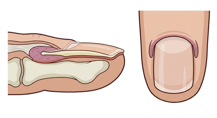
The terminology is hard to follow without a picture, and several of the steps below refer to structures that are not visible when looking at the nail.
- The nail plate is the hard, visible nail.
- The nail bed is the tissue beneath the nail plate; a laceration here heals with a deformed nail unless it is sutured.
- The matrix lies most proximally, largely hidden beneath the proximal nail fold, and is the only tissue that produces new nail. Its visible distal part is the lunula.
- The matrix horns are the lateral extensions of the matrix, reaching out to the sides beneath the nail folds. It is these that must be destroyed during matrix ablation — miss the horn and the nail spike returns in the same corner.
- The nail folds are the skin folds proximal and lateral to the plate, and the cuticle is the flap of skin that extends from the proximal fold over the base of the plate.
Preparation and equipment
- Chlorhexidine in alcohol or an iodine solution.
- Lidocaine 10 mg/mL without adrenaline for the block (adrenaline is safe in fingers and toes according to modern data, but there is rarely any reason to use it when a tourniquet is being applied anyway), a 27 G needle, a 5 mL syringe.
- A tourniquet: a Penrose drain with artery forceps, or the cut-off finger of a sterile glove rolled down.
- A nail splitter or strong pointed scissors, a Freer elevator or small artery forceps, toothed surgical forceps.
- A size 11 scalpel.
- A chemical cautery agent: phenol 88 %, sodium hydroxide 10 % or silver nitrate.
- Electrocautery or a heated paper clip for trephination.
- Antibiotic ointment, a non-adherent dressing, gauze.
The digital block
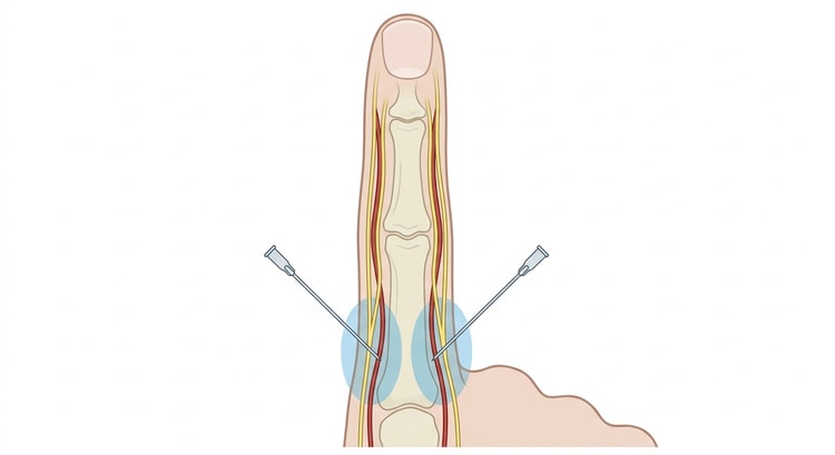
Anaesthetise from the dorsal aspect, one injection medial and one lateral to the base of the phalanx, 2–3 mL on each side, with the needle tip directed volarly until a weal forms over the volar nerve. Wait 10 minutes — insufficient waiting time is the commonest reason for the patient finding the procedure painful. Test with a needle tip before you start.
Apply the tourniquet only after the block has taken effect, and note the time. It must not stay on for longer than 20–30 minutes.
Procedure
Paronychia — incision and drainage
- A digital block if the abscess is more than superficial.
- Introduce a Freer elevator, small artery forceps or the back of a size 11 scalpel blade between the nail and the nail fold, parallel to the nail, and lift the fold off the nail plate.
- A skin incision is usually not needed. With a large abscess pointing through the skin, an incision can be made parallel to the nail fold.
- Express the pus and send it for culture.
- If necessary, place a thin gauze wick under the fold for 24–48 hours.
- If the abscess extends beneath the nail plate (subungual extension), the proximal or lateral part of the nail must be removed.
Antibiotics are given for surrounding cellulitis, immunosuppression or diabetes; the first choice is flucloxacillin, with anaerobic cover added after nail biting or oral contact.
Ingrown nail — splinting
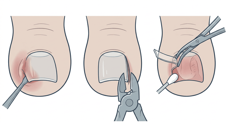
Lift the lateral edge of the nail with artery forceps and slide a cotton wisp, a strip of suture material or a 1 cm length of a 2–3 mm drain beneath the edge with a sawing movement until the nail lifts free of the fold. It is changed daily after washing and removed once the inflammation has settled and the nail has grown beyond the fold.
Ingrown nail — partial avulsion
- A digital block and a tourniquet.
- Free the lateral 3–4 mm of the nail plate from the nail bed with an elevator, all the way in beneath the cuticle to the matrix.
- Cut the nail longitudinally with a nail splitter, all the way to the base. If no splitter is available, use strong pointed scissors with upward pressure to spare the nail bed.
- Grasp the segment with artery forceps close to the root and twist it away from the nail bed until it comes free from the fold and the cuticle.
- Inspect to ensure that no spicules remain.
- In recurrent disease: destroy the corresponding part of the matrix with phenol 88 % on a cotton bud for 30 seconds × 3, drying between applications, and then irrigate copiously with saline or alcohol. Phenol must not come into contact with the surrounding skin.
- Release the tourniquet, check the circulation, apply antibiotic ointment and a non-adherent dressing.
Subungual haematoma — trephination
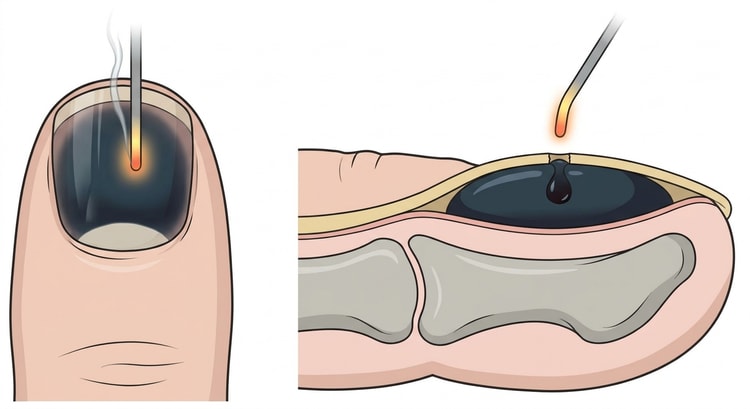
Burn a hole through the nail plate with electrocautery or a red-hot paper clip, in the middle of the haematoma. The nail plate has no nerve supply; a block is rarely needed. Hold it in place until the blood has drained. With a concomitant fracture of the distal phalanx, trephination is still indicated, but the wound must then be regarded as an open fracture.
The dividing line lies at the nail plate, not at the size of the haematoma. The older rule that a haematoma involving more than 25 or 50 % of the nail surface requires nail avulsion and suture of the nail bed has not held up in follow-up studies: as long as the nail plate and the edges of the nail folds are intact, the appearance of the nail is just as good after trephination alone, whatever the extent of the haematoma and whether or not there is an underlying tuft fracture. Nail avulsion with inspection and suture of the nail bed is, however, indicated when
- the nail plate is split, partly loosened or avulsed,
- the edge of the nail fold is torn or the base of the nail plate lies on top of the nail fold,
- there is a displaced fracture of the terminal phalanx requiring reduction.
Complications
- Infection, at worst osteomyelitis of the distal phalanx — particularly in patients with diabetes.
- Nail bed injury with permanent nail deformity.
- Recurrence of the ingrown nail (10–30 % without matrix ablation, under 5 % with phenol).
- An inclusion cyst in the nail fold.
- Hyperkeratosis, oedema and persistent redness of the nail fold.
- Digital ischaemia from a forgotten tourniquet — count the instruments and the tourniquet at the end.
- Chemical burns of the surrounding skin from spilled phenol.
Aftercare and follow-up
Antibiotic ointment and a non-adherent dressing. After partial avulsion: soaks in lukewarm water 2–3 times daily from day one, with a daily change of dressing. Elevation and rest for the first 24 hours. Roomy shoes.
Inspect after 2–3 days with infection in mind. Phenol-treated wounds often weep for 2–4 weeks — this is expected and is not in itself a sign of infection.
Give instructions on cutting the nail straight across, not rounded at the corners, and on shoes that do not press. In patients with diabetes: refer for specialist foot care.
Common pitfalls
- Insufficient waiting time after the block — always test with a needle tip.
- No tourniquet, which gives a blood-filled field and spicules left behind.
- The nail not cut all the way to the base — the single commonest cause of recurrence.
- Incising a herpetic whitlow. Grouped vesicles and severe pain without pus — treat with aciclovir, do not incise.
- A missed subungual melanoma behind a "haematoma" that does not grow out with the nail.
- Nail avulsion merely because the haematoma is large. If the nail plate and the edge of the fold are intact, trephination is enough; if they are damaged, it is not enough however small the haematoma.
- Phenol on healthy skin, or inadequate irrigation afterwards.
- A diabetic foot without a vascular assessment — the procedure can start a chronic ulcer.

