
The ankle-brachial index is the cheapest and most accessible method of objectively demonstrating peripheral arterial disease, and it is needed more often than it is used — not only in claudication, but before every compression treatment of a leg ulcer and as a risk marker in diabetes and known atherosclerotic disease. An ABI ≤ 0.90 is diagnostic of peripheral arterial disease and at the same time indicates a substantially increased risk of myocardial infarction and stroke.
Indications
- Intermittent claudication, rest pain in the foot or leg, or a non-healing distal ulcer.
- Before compression treatment of a leg ulcer — compression in arterial insufficiency can cause necrosis.
- Screening in diabetes of > 10 years' duration, or in diabetes with age > 50 years.
- Risk assessment in known coronary or cerebrovascular disease.
- Follow-up after vascular surgery or endovascular intervention.
- Assessment of healing potential in ulcers and before deciding the level of amputation.
Contraindications
There are no absolute contraindications. Refrain or modify in:
- Deep vein thrombosis in the leg — cuff compression should be avoided in the acute phase.
- A leg that has recently been operated on, is in a cast or has extensive wounds so that the cuff cannot be applied. Place the cuff over a protective dressing, or measure at the toe instead.
- Marked pain that prevents the patient from tolerating compression.
Preparation and equipment
Equipment: a handheld Doppler probe 5–10 MHz, ultrasound gel, a blood pressure cuff of the correct size (the width should correspond to about 40 % of the circumference of the limb), a manual manometer, plastic film or a dressing to protect an ulcer, and where needed a photoplethysmograph and a toe cuff.
Preparation:
- The patient must have rested lying down for at least 10 minutes in a room at a comfortable temperature — a cold foot gives falsely low pressures.
- No smoking or coffee in the preceding hour.
- Remove socks and any compression stockings.
- Avoid measuring the arm pressure on the side with a dialysis fistula, lymphoedema after axillary clearance or a recently inserted venous port.
Procedure
- Arm pressure. Place the cuff on the upper arm. Locate the brachial signal in the antecubital fossa with the Doppler probe at 45–60° to the skin. Inflate to 20 mmHg above the point at which the signal disappears, deflate slowly (2 mmHg/s) and note the pressure at which the signal returns. Repeat on the other arm.
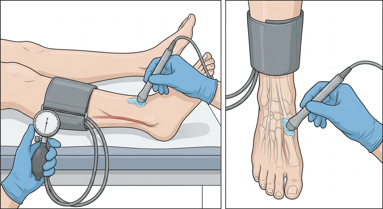
- Ankle pressure. Place the cuff just above the malleoli, with its lower edge about 2 cm above the tip of the malleolus.
- Dorsalis pedis artery — the signal is sought on the dorsum of the foot lateral to the extensor hallucis longus tendon. Measure in the same way as on the arm.
- Posterior tibial artery — behind the medial malleolus. Measure.
- Repeat on the other leg. If the signal is uncertain or borderline, the fibular artery can also be sought anterior to the lateral malleolus.
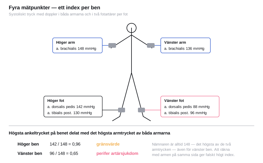
Calculation: for each leg, the highest of the dorsalis pedis and posterior tibial pressures is divided by the highest arm pressure of the two arms. Each leg gets its own index.
ABI = highest ankle pressure in that leg ÷ highest arm pressure of the two arms
Interpretation
| ABI | Interpretation | Action |
|---|---|---|
| > 1.40 | Incompressible vessels — medial sclerosis | Measure the toe pressure; the ABI is not usable |
| 1.00–1.40 | Normal | If suspicion persists: exercise ABI |
| 0.91–0.99 | Borderline | Exercise ABI or follow-up |
| 0.41–0.90 | Peripheral arterial disease, mild to moderate | Secondary prevention, walking exercise |
| ≤ 0.40 | Severe ischaemia | Urgent vascular surgical assessment |
flowchart TD
  A[ABI calculated for each leg] --> B{Index above 1.40 or an incompressible signal?}
  B -- Yes --> C[Medial sclerosis: the value cannot be interpreted]
  C --> D[Measure the toe pressure and the toe-brachial index]
  D --> E{Toe-brachial index at least 0.70?}
  E -- Yes --> F[Peripheral arterial disease unlikely]
  E -- No --> G[Peripheral arterial disease is present despite a high ABI]
  B -- No --> H{Index}
  H -- At least 1.00 --> I[Normal: exercise ABI in typical claudication]
  H -- 0.91 to 0.99 --> J[Borderline: exercise ABI or follow-up]
  H -- 0.41 to 0.90 --> K[Peripheral arterial disease: secondary prevention and walking exercise]
  H -- At most 0.40 --> L[Severe ischaemia: urgent vascular surgical assessment]
Exercise ABI. With a normal or borderline resting ABI but typical claudication, the ankle pressure is measured immediately after treadmill walking or after repeated heel raises. A fall of ≥ 20 % after exercise confirms peripheral arterial disease.
Toe-brachial index (TBI). The digital arteries rarely calcify, which makes toe pressure the method of choice in diabetes and renal failure. A normal TBI is ≥ 0.70. A toe pressure < 30 mmHg indicates poor healing potential; > 60 mmHg indicates good healing potential.
Segmental pressures along the thigh and calf localise the obstruction to the aortoiliac, femoropopliteal or crural level, and are measured in a vascular laboratory.
Complications
The investigation is in practice free of risk. Occasional patients find the cuff uncomfortable, and in marked ischaemia the compression can be painful. In a patient with an ulcer the cuff must be placed over a clean protective layer to avoid contamination.
Aftercare and follow-up
An abnormal ABI is not just a diagnosis of the leg — it is a systemic risk marker. Whatever the symptoms, the patient should receive smoking cessation, a statin, blood pressure and diabetes control, and antiplatelet therapy. Structured walking exercise is the first-line treatment in claudication.
| Finding | Management |
|---|---|
| ABI 0.41–0.90 with claudication | Secondary prevention + walking exercise; referral if the effect is insufficient after 3 months |
| ABI ≤ 0.40 or rest pain | Vascular surgical assessment, prioritised |
| Non-healing ulcer with ABI < 0.80 | Vascular assessment before compression treatment |
| ABI > 1.40 | Toe pressure, supplemented where needed by duplex ultrasound |
Compression treatment of venous leg ulcers requires an ABI ≥ 0.80 for full compression (40 mmHg at the ankle); at 0.50–0.79 reduced compression is used, about 20–30 mmHg, under vascular surgical supervision, and below 0.50 compression must not be given. During ongoing compression treatment the ABI is measured about every three to six months, and always with new or increasing pain in the leg.
Documentation
An index without the underlying pressures cannot be evaluated afterwards. Record in the notes:
- All measured pressures: both arms, and the dorsalis pedis and posterior tibial arteries in each foot, in mmHg.
- Which vessel gave the pressure used in the numerator for each leg.
- The index for each leg to two decimal places, and which arm pressure formed the denominator.
- Signal quality and waveform if the equipment displays it — a triphasic, biphasic or monophasic signal tells you about the state of the vessel even when the pressure is normal.
- The resting time before the measurement, and any vessel that could not be located.
Common pitfalls
- A falsely normal or high ABI in diabetes and renal failure because of medial sclerosis — measure the toe pressure instead.
- The wrong reference arm. Use the higher of the two arm pressures; otherwise a subclavian stenosis on one side makes the index falsely high.
- Too short a period of rest before the measurement gives raised pressures and thus an incorrect index.
- The wrong cuff width — a cuff that is too narrow overestimates the pressure.
- Palpation instead of Doppler. A palpable pulse does not exclude peripheral arterial disease, and palpation-based pressure measurement systematically underestimates.
- Deflating too rapidly when reading, which misses the point of return by several mmHg.
- A normal resting ABI interpreted as a normal vascular status in typical claudication — add an exercise ABI.

