
The value of the exercise test lies not in ST depression alone but in the overall picture: exercise capacity, symptoms, blood pressure response, heart rate response, arrhythmias and recovery. A patient who stops at a low workload with a fall in blood pressure is a high-risk patient even with normal ST segments, whereas isolated ST depression in a woman with a low pre-test probability is usually a false positive.
Indications
- Evaluation of chest pain with an intermediate pre-test probability of coronary artery disease.
- Assessment of exercise capacity and symptoms in known heart disease.
- Risk assessment after myocardial infarction or before revascularisation.
- Evaluation of treatment effect in stable angina.
- Exercise-induced arrhythmia or exercise-induced syncope.
- Assessment of chronotropic function and pacemaker settings.
- Preoperative functional assessment before major surgery.
At a low pre-test probability most positive results are false positives; at a high probability a negative test adds little. Imaging (coronary CT angiography, myocardial perfusion scintigraphy, stress echocardiography) has greater diagnostic accuracy.
Contraindications
Absolute:
- Acute coronary syndrome within 48 hours, or ongoing unstable angina.
- Uncontrolled symptomatic arrhythmia.
- Symptomatic severe aortic stenosis.
- Decompensated heart failure.
- Acute pulmonary embolism or deep vein thrombosis.
- Acute myocarditis, pericarditis or endocarditis.
- Acute aortic dissection.
- Physical inability to perform the test safely.
Relative:
| Condition | Comment |
|---|---|
| Left main stenosis | Known — perform the test only on a clear indication and with full preparedness |
| Moderate to severe aortic stenosis without symptoms | Individual assessment |
| Hypertrophic cardiomyopathy with outflow tract obstruction | Caution |
| Tachyarrhythmia or bradyarrhythmia | Rate control first |
| Blood pressure > 200/110 mmHg at rest | Treat first |
| Electrolyte disturbance | Correct |
| Second- or third-degree AV block | Depending on the indication |
| Marked anaemia | Correct |
A resting ECG that makes ST interpretation impossible — left bundle branch block, paced rhythm, pre-excitation, digitalis effect, marked LVH with ST-T changes — is an argument for imaging rather than a conventional exercise test.
Preparation and equipment
- Cycle ergometer with a braked, workload-controlled resistance; 12-lead ECG with continuous recording; automated or manual blood pressure measurement.
- Resuscitation trolley with defibrillator, oxygen, suction and emergency drugs in the room.
- Staff with current CPR competence; a physician available.
Patient preparation:
- A light meal is permitted; no large meal, no coffee and no nicotine in the preceding 2–3 hours.
- Comfortable clothing and shoes.
- Beta blockers: withheld 2–3 days before the test only when the question is purely diagnostic and the target heart rate is needed for the test to be conclusive. For assessment of function and prognosis, arrhythmia evaluation, known coronary artery disease and evaluation of treatment effect, the medication is continued — abrupt withdrawal carries a risk of rebound. The referring physician should have made this decision; document which applies.
- Nitroglycerin is continued; note it in the report.
- Shaving and careful skin preparation at the electrode sites — movement artefact ruins an exercise test more often than anything else.
Record a resting ECG both supine and sitting, and resting blood pressure in both arms.
Procedure
- Exercise protocol: start at 25–50 W and increase by 10–25 W every minute, or by 25–50 W every third minute. Adjust so that the total exercise time is 8–12 minutes — a shorter test gives insufficient information, a longer one is limited by local muscle fatigue rather than by the heart.
- Pedalling rate 50–70 revolutions per minute, kept steady.
- ECG continuously, with a printout every minute and on symptoms.
- Blood pressure every second minute and on symptoms.
- Ask actively about chest pain, breathlessness and leg fatigue; have the patient rate exertion on the Borg RPE scale 6–20.
- Encourage maximal effort. Target heart rate may be estimated as 220 − age, but the test should be driven to symptom limitation, not to a number.
- Recovery: continue recording ECG and blood pressure for at least 6 minutes after exercise ends, with the patient seated. Most ST depression appears or persists here.
Termination criteria
flowchart TD
  A[Ongoing exercise, continuous ECG and blood pressure every second minute] --> B{ST elevation ≥ 1 mm in a lead without a Q wave, except aVR, aVL and V1?}
  B -- Yes --> Z[Stop immediately]
  B -- No --> C{Fall in systolic blood pressure > 10 mmHg with rising workload?}
  C -- Yes, with other signs of ischaemia --> Z
  C -- Yes, without other signs of ischaemia --> Y[Relative criterion - weigh against the indication]
  C -- No --> D{Sustained ventricular tachycardia, second- or third-degree AV block with haemodynamic compromise, or moderate to severe angina?}
  D -- Yes --> Z
  D -- No --> E{Cyanosis, pallor, cold sweating, ataxia, dizziness or presyncope?}
  E -- Yes --> Z
  E -- No --> F{Marked ST depression, hypertensive response, other arrhythmia, new bundle branch block, severe dyspnoea or claudication?}
  F -- Yes --> Y
  F -- No --> G{Does the patient want to stop, or is the patient symptom-limited?}
  G -- Yes --> H[End the test as planned and continue recording during recovery]
  G -- No --> A
  Z --> H
  Y --> H
Absolute:
- ST elevation ≥ 1 mm in a lead without a Q wave (except aVR, aVL and V1).
- A fall in systolic blood pressure > 10 mmHg during rising workload, with other signs of ischaemia.
- Moderate to severe angina.
- Sustained ventricular tachycardia.
- Second- or third-degree AV block appearing during exercise and compromising haemodynamics.
- Signs of impaired perfusion: cyanosis, pallor, cold sweating.
- Neurological symptoms, ataxia, dizziness, presyncope.
- Monitoring technically impossible.
- The patient wants to stop.
Relative: marked ST depression (> 2 mm horizontal or downsloping), a fall in blood pressure without other signs of ischaemia, increasing chest pain, an arrhythmia that is not sustained VT, bundle branch block appearing during exercise that cannot be distinguished from VT, a hypertensive response (systolic
250 mmHg and/or diastolic > 115 mmHg), severe dyspnoea or claudication.
The list follows the AHA Exercise Standards. Swedish laboratories apply their own, often stricter, standard operating procedures — several regions terminate at a systolic pressure ≥ 280 mmHg, at a systolic fall ≥ 15 mmHg on a single measurement or ≥ 10 mmHg on repeated measurements, and at ST depression ≥ 3–4 mm. Follow the local standard operating procedure and do not mix the numbers.
Interpretation
| Parameter | Pathological finding |
|---|---|
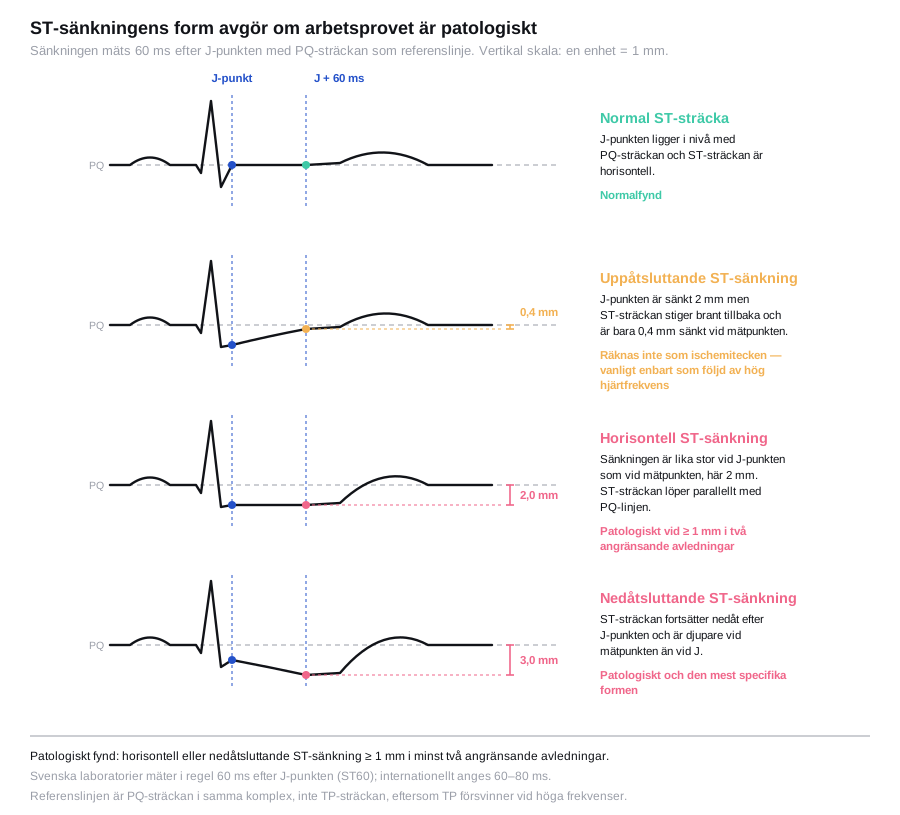
| ST depression | ≥ 1 mm horizontal or downsloping, measured 60 ms after the J point, in two contiguous leads |
| Upsloping ST depression | Does not count as a sign of ischaemia. Only slowly upsloping depression ≥ 1.5–2 mm is equivocal, with low specificity |
| ST elevation | ≥ 1 mm in a lead without a Q wave — transmural ischaemia, high risk |
| Blood pressure | Failure to rise, or a fall > 10 mmHg during rising workload |
| Exercise capacity | < 5 metabolic equivalents, or < 85 % of that predicted for sex and age |
| Heart rate | Chronotropic incompetence: < 80 % of the predicted heart rate reserve |
| Recovery heart rate | A fall < 12 beats in the first minute with an active cool-down in the upright position (< 18 beats if the patient is laid down immediately without a cool-down) |
| Arrhythmia | VT or frequent ventricular ectopic beats during exercise and in recovery |
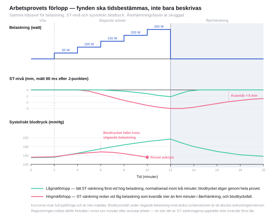
High risk is suggested by: ST depression at a low workload, ST depression in many leads, ST depression persisting for more than five minutes into recovery, a fall in blood pressure, ST elevation, low exercise capacity, and VT.
State in the report where in the course the findings appeared, at what workload and heart rate, and which symptoms were present. "Positive exercise test" without contextual information is not a useful report.
Sensitivity is around 60–70 % and specificity 70–80 %, but sensitivity varies widely with the extent of disease: below 50 % in single-vessel disease and around 85 % in three-vessel or left main disease. False positive results are more common in women, in left ventricular hypertrophy, digitalis treatment, hypokalaemia and in the presence of resting ST changes.
Complications
Serious events occur in approximately 1 per 10,000 examinations: myocardial infarction, sustained ventricular arrhythmia, cardiac arrest and death. More common but less serious are vasovagal reactions, musculoskeletal pain, dizziness and a marked hypertensive reaction. It is the preparedness in the room, not the statistics, that determines the outcome when it happens.
Aftercare and follow-up
Let the patient remain seated under observation until heart rate, blood pressure and ECG have normalised, for at least 6 minutes, and longer if there are symptoms or persisting changes. Beta blockers that were withheld are restarted the same day.
| Result | Management |
|---|---|
| Normal test, good exercise capacity | Low risk; consider another cause of the symptoms |
| Pathological with a low-risk profile | Optimised medical therapy, clinical follow-up |
| High-risk findings | Prompt contact with cardiology, often coronary angiography |
| Inconclusive (target heart rate not reached, artefact) | Complete the evaluation with imaging |
Common pitfalls
- The test is stopped too early at a submaximal workload and reported as negative — that is inconclusive, not negative.
- Too rapid an increase in workload means the legs give up before the heart has been stressed.
- Beta blockers continued for a diagnostic test make the target heart rate unattainable.
- The recovery phase is cut short. A large proportion of ST depression appears only there.
- ST interpretation on an ECG that cannot be interpreted — left bundle branch block, paced rhythm or pre-excitation.
- An exercise test in a patient with a low pre-test probability generates false positive results and unnecessary investigations.
- A report without context. Workload, heart rate, symptoms and time course are part of the finding.


