
Two recordings that are performed in primary care but interpreted as specialist investigations. Both fail more often for practical than for medical reasons: the cuff is in the wrong place, the electrodes come off, the patient has not kept a diary. The set-up and the instructions decide whether the investigation will be usable.
Ambulatory blood pressure monitoring (ABPM)
Indications
- Confirmation of newly detected hypertension — recommended before treatment is started.
- Suspected white-coat hypertension: raised office blood pressure without organ damage.
- Suspected masked hypertension: normal office blood pressure but organ damage or high risk.
- Treatment-resistant hypertension.
- Suspected nocturnal hypertension or absent nocturnal dipping (diabetes, renal failure, sleep apnoea).
- Suspected hypotension: dizziness, syncope, autonomic dysfunction, adverse drug effects.
- Wide variability between measurements.
- Hypertension in pregnancy.
Contraindications
There are no absolute contraindications. Refrain or adapt in atrial fibrillation with a rapid and irregular ventricular rate (oscillometric measurement becomes unreliable), lymphoedema or a dialysis fistula in both arms, an arm circumference too large for the available cuffs, and in a patient who does not tolerate repeated compression (for example marked thrombocytopenia).
Set-up and instructions
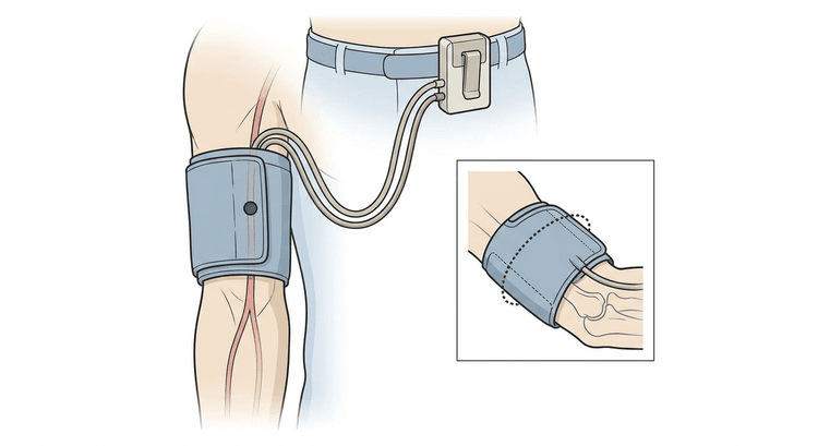
- Choose the cuff according to arm circumference; a cuff that is too narrow overestimates the pressure. Measure the circumference, do not guess.
- Measure the blood pressure in both arms when fitting the device. Use the arm with the higher pressure, or the non-dominant arm if the values are equal. Check against a manual measurement and document the difference.
- Programme measurements every 20 minutes during the day and every 30 minutes at night, or alternatively every 15 and every 30 minutes respectively.
- Instruct the patient to stop, keep the arm still and lowered at each measurement, and not to talk.
- Otherwise ordinary activities, including work. No showering or bathing.
- A diary of going to bed, getting up, medication intake, symptoms and any unusual activities.
Interpretation
A recording is acceptable if at least 70 % of the measurements are valid, with at least 20 daytime and 7 night-time values, and with a measurement interval of no more than 30 minutes throughout the 24 hours.
| Phenotype | Office blood pressure | ABPM |
|---|---|---|
| Normotension | Normal | Normal |
| White-coat hypertension | Raised | Normal |
| Masked hypertension | Normal | Raised |
| Established hypertension | Raised | Raised |
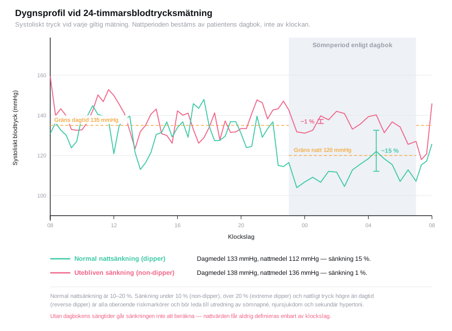
Nocturnal dipping: the pressure normally falls by 10–20 % during the night.
| Pattern | Nocturnal fall | Comment |
|---|---|---|
| Dipper | 10–20 % | Normal |
| Non-dipper | < 10 % | Independent risk marker |
| Reverse dipper | ≤ 0 %, night-time pressure higher than daytime | The highest risk in the group |
| Extreme dipper | > 20 % | Also unfavourable, particularly in the elderly |
An abnormal 24-hour rhythm should prompt investigation for sleep apnoea, renal disease and secondary hypertension, and lead to one dose being moved to the evening. The fall can only be calculated if the diary states when the patient fell asleep and got up.
Holter ECG and longer rhythm monitoring
Indications
- Palpitations, presyncope or syncope of suspected arrhythmic origin.
- Assessment of rate control in atrial fibrillation.
- Suspected paroxysmal atrial fibrillation, including after cryptogenic stroke.
- Evaluation of antiarrhythmic treatment or pacemaker function.
- Suspected bradyarrhythmia or pauses, particularly at night.
- Symptom–rhythm correlation in non-specific complaints.
The choice of recording is governed by the frequency of symptoms:
| Symptom interval | Method |
|---|---|
| Daily | 24-hour Holter |
| A few times per week | 48–72-hour Holter or 7-day recording |
| About once a month | Thumb ECG or event recorder, 2–4 weeks |
| Less often, with syncope | Implantable loop recorder |
Contraindications
None. Practical obstacles are skin disease or allergy to the electrode gel, and marked body hair that cannot be shaved.
Set-up
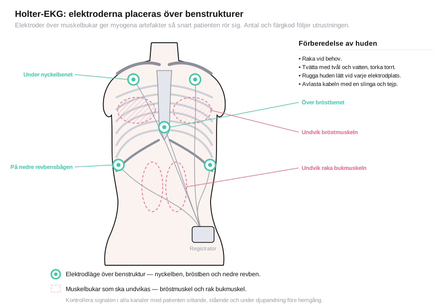
- Shave, wash with soap and water, dry and lightly abrade the skin at each electrode site. Grease and skin scales are the commonest cause of unusable recordings.
- Place the electrodes over bony structures — sternum, ribs, clavicle — not over muscle bellies, in order to reduce myogenic artefacts.
- Relieve strain on the cables with a loop and tape, so that traction does not reach the electrode.
- Check the signal quality in all channels before the patient goes home, preferably with the patient sitting, standing and taking deep breaths.
- Give a symptom diary and explain the event button: press it at the time of symptoms, note the time and what was being done. Without a diary, symptoms and rhythm cannot be linked.
- No showering. Otherwise normal activity.
Interpretation
flowchart TD
  A[The recording read together with the symptom diary] --> B{Did the patient have their typical symptoms during the recording?}
  B -- No --> C[The question is unanswered - extend the recording or change method, do not dismiss the suspicion of arrhythmia]
  B -- Yes --> D{Was there a concurrent arrhythmia at the time of symptoms?}
  D -- Yes --> E[Symptom-rhythm correlation demonstrated - treat the arrhythmia]
  D -- No --> F[A valuable negative finding - argues against an arrhythmic cause, look for another cause]
  A --> G{Findings without concurrent symptoms?}
  G -- Pauses over 3 seconds during the day, Mobitz II or third-degree AV block --> H[Bradyarrhythmia workup, assess for pacemaker indication]
  G -- Episodes of atrial fibrillation --> I[State the AF burden and the ventricular rate, assess the stroke risk]
G -- PVC burden 10 percent or more, or non-sustained VT --> J[Echocardiography and further cardiological workup]| Finding | Comment |
|---|---|
| Sinus bradycardia down to 30–40/min at night | Normal in well-trained and young people |
| Pauses < 3 seconds at night | Often normal; > 3 s during the day requires investigation |
| Occasional PVCs and PACs | Common; a PVC burden ≥ 10 % raises suspicion of PVC-induced cardiomyopathy and warrants echocardiography. The risk rises markedly above 20 %; there is no sharp threshold |
| Non-sustained VT | Investigate for underlying structural heart disease |
| Episodes of atrial fibrillation | State the total AF burden and the ventricular rate |
| Mobitz II or third-degree AV block | Assess for pacemaker indication |
| Symptoms without concurrent arrhythmia | A valuable negative finding — argues against an arrhythmic cause |
A Holter ECG without symptoms during the recording does not answer the question. Always state whether the patient had their typical complaints during the recording period.
Complications
Contact dermatitis and skin irritation from the electrodes or the cuff; petechiae and tenderness in the arm after repeated compressions; disturbed sleep from night-time measurements. Serious complications do not occur.
Aftercare and follow-up
Remove the equipment, download the recording, and go through the diary together with the recording — that is where the interpretation is made. Inform the patient of the time to a result.
For ABPM: start or adjust treatment according to the 24-hour values, not according to the office blood pressure. White-coat hypertension is as a rule not treated pharmacologically but must be followed up — a substantial proportion go on to develop established hypertension. Repeat ABPM after 1–2 years.
For Holter: a negative recording with persisting symptoms warrants a longer recording, not dismissal of the question.
Common pitfalls
- The wrong cuff size — the commonest cause of erroneous ABPM results.
- No diary. Without sleep times the 24-hour rhythm cannot be calculated, and without notes on symptoms the Holter cannot be correlated.
- Inadequate skin preparation produces artefacts that make the recording uninterpretable.
- Too few valid measurements being reported anyway — check the proportion of valid values before the report is written.
- ABPM in atrial fibrillation gives unreliable oscillometric values.
- A short Holter recording for infrequent symptoms. Choose the recording time according to the frequency of symptoms.
- Interpreting night-time values as abnormal without knowing when the patient actually slept.

