
Compression is the treatment that heals venous leg ulcers; everything else is supportive. But the same compression applied to a limb with arterial insufficiency can cause necrosis and, in the worst case, amputation. The decisive step is therefore not the choice of dressing but the ankle-brachial index before the first bandage is applied.
Indications
- Venous leg ulcer — about 70% of all leg ulcers.
- Chronic venous oedema, lipodermatosclerosis, stasis dermatitis.
- Prevention of recurrence after a healed venous ulcer.
- After deep vein thrombosis with post-thrombotic syndrome.
Differential diagnosis
| Feature | Venous ulcer | Arterial ulcer | Diabetic foot ulcer |
|---|---|---|---|
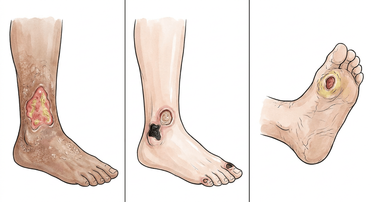
| Site | Gaiter area, above the medial malleolus | Toes, heel, lateral malleolus, pressure points | Beneath the metatarsal heads, toes, heel |
| Edge | Irregular, shallow | Sharp, punched-out | Surrounded by callus |
| Wound bed | Fibrin-covered, granulating, copious exudate | Dry, necrotic, pale | Variable, often deep |
| Pain | Moderate, relieved by elevation | Severe, worse on elevation, better when dependent | Often painless (neuropathy) |
| Surrounding skin | Hyperpigmentation, oedema, lipodermatosclerosis, eczema | Pale, hairless, atrophic, cold | Dry, hyperkeratotic |
| Pulses | Palpable | Weak or absent | May be palpable despite ischaemia |
| ABI | > 0.9 | < 0.9 | Often falsely high |
Contraindications to compression
The ankle-brachial index measured with Doppler determines whether compression may be applied at all, and if so at what pressure. Palpable foot pulses are not an acceptable substitute, and in patients with diabetes or renal failure the index may be falsely normal because of medial sclerosis — in that case add toe pressure measurement.
flowchart TD
  A[Leg ulcer] --> B[Measure the ankle-brachial index with Doppler before the first bandage]
  B --> C{Index}
  C -- At least 0.80 --> D[Full compression, 40 mmHg at the ankle]
  C -- 0.50 to 0.79 --> E[Reduced compression, 20 to 30 mmHg]
  E --> F[Vascular surgical assessment in parallel]
  C -- Below 0.50 --> G[Compression contraindicated]
  G --> H[Urgent vascular surgical assessment]
  C -- Above 1.40 or diabetes with suspected medial sclerosis --> I[The index cannot be interpreted: measure toe pressure]
  I --> J{Toe pressure}
  J -- Below 30 mmHg --> G
  J -- At least 30 mmHg --> E
  D --> K[Reassess the index every three to six months and on any new pain]
  E --> K
Absolute:
- ABI < 0.5 or toe pressure < 30 mmHg.
- Decompensated heart failure — compression of both legs acutely increases preload.
- Untreated deep vein thrombosis in the acute phase.
- Septic phlebitis, ongoing soft tissue infection with systemic involvement.
Relative:
| Condition | Adaptation |
|---|---|
| ABI 0.5–0.79 | Reduced compression (20–30 mmHg) under vascular surgical supervision |
| Diabetes with neuropathy | Reduced compression, closer monitoring — the patient cannot feel pressure injury |
| Peripheral neuropathy of other cause | As above |
| Severe pain | Titrate the pressure up gradually |
| Rheumatoid arthritis with vasculitis | Individual assessment |
Repeat the ABI every three to six months during ongoing compression therapy, and always in the event of new or increasing pain.
Preparation and equipment
- Doppler for the ankle-brachial index, blood pressure cuff.
- Lukewarm tap water or saline for cleansing, swabs.
- Sharp spoon (curette), ring curette, toothed forceps, scissors, no. 15 scalpel.
- Topical anaesthetic cream (lidocaine–prilocaine) or infiltration anaesthesia for painful debridement.
- Wound bed dressing chosen by exudate volume, edge protection (zinc paste, barrier film).
- Compression materials: two-layer system, four-layer system, short-stretch bandage, zinc paste bandage (Unna boot), or a class 2–3 compression stocking.
- Tape measure for ankle circumference — this governs the choice of material and bandaging technique.
Procedure
Assessment
- History: duration, previous ulcers, DVT, varicose veins, claudication, diabetes, smoking, character of the pain.
- Measure and trace the ulcer; photograph it with a ruler in the image. A reduction in size of 40% within four weeks predicts healing — failure to shrink should prompt reconsideration of the diagnosis.
- Palpate the pulses and measure the ankle-brachial index with Doppler.
- Culture only in the presence of clinical signs of infection — all chronic wounds are colonised, and a culture taken without clinical suspicion leads only to unnecessary antibiotics.
- Biopsy any ulcer that has not healed within three months despite adequate treatment — malignancy (Marjolin's ulcer, basal cell carcinoma) and vasculitis do occur.
Wound debridement
- Cleanse with lukewarm tap water or saline; showering the leg is a good option.
- Apply topical anaesthetic cream under occlusion for 30–45 minutes before painful debridement.
- Remove fibrin, necrosis and callus from the edges with a curette or sharp spoon, using even tangential strokes until granulation tissue or pinpoint bleeding appears.
- Avoid damaging granulation tissue. Stop in the event of heavy bleeding or unacceptable pain — debridement can be divided over several visits.
- Alternatives in the presence of pain or suspected ischaemia: autolytic debridement with hydrogel, enzymatic debridement, or larval therapy.
- Do not debride dry, stable necrosis on an ischaemic limb — it acts as a biological barrier until the circulation has been addressed.
Choice of wound dressing
| Wound bed | Dressing |
|---|---|
| Copious exudate | Foam, alginate, hydrofibre |
| Moderate exudate | Foam |
| Dry wound bed | Hydrogel, hydrocolloid |
| Fibrin-covered | Hydrogel or hydrofibre under compression |
| Clinically infected | Silver- or iodine-containing dressing, short term; systemic antibiotics for cellulitis |
| Granulating | Non-adherent, change infrequently |
The choice of dressing affects healing time only marginally compared with compression. Choose according to exudate volume and change interval, not by product name. Protect the wound edges from maceration with zinc paste or a barrier film.
Compression bandaging
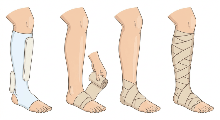
The target is 40 mmHg at the ankle for a venous ulcer, with pressure decreasing gradually proximally. The bandage is applied in the morning, before the leg has swollen.
- The foot is held in 90° dorsiflexion throughout bandaging.
- Pad behind the malleoli, over the tendon and along the tibial crest — this is where pressure injuries arise.
- Start at the base of the toes, include the heel, and bandage upwards to just below the popliteal fossa.
- Use 50% overlap and even tension according to the manufacturer's markings.
- If the ankle circumference is below 18 cm, additional padding is required to achieve the correct pressure without pressure necrosis.
- Check the toes: colour, temperature and capillary refill immediately after bandaging.
| System | Change interval | Comment |
|---|---|---|
| Four-layer system | Once weekly | High, sustained pressure; requires trained staff |
| Two-layer system | Once weekly | Thinner, fits inside ordinary shoes |
| Short-stretch bandage | Daily or every other day | High working pressure, low resting pressure — useful when the circulation is impaired |
| Zinc paste bandage (Unna boot) | Once weekly | Also treats stasis dermatitis |
| Class 2–3 compression stocking | Daily | Prevention of recurrence after healing; requires that the patient can put it on |
Levels of compression. The class denotes the pressure at the ankle, and the pressure must always decrease proximally. The prescription should state both the level and the ankle-brachial index on which it rests.
| Class | Pressure at the ankle | Use |
|---|---|---|
| Class 1 | 18–21 mmHg | Mild oedema, superficial varicose veins; may be used at ABI 0.50–0.79 when higher pressure is not tolerated |
| Class 2 | 23–32 mmHg | Venous insufficiency, after a healed venous ulcer, after DVT; corresponds to reduced compression |
| Class 3 | 34–46 mmHg | Marked oedema and active venous ulcer — the level corresponding to the 40 mmHg target |
| Class 4 | > 49 mmHg | Lymphoedema; not relevant for leg ulcers in primary care |
Compression is contraindicated at an ABI < 0.50 or a toe pressure < 30 mmHg, and at an ABI of 0.50–0.79 the pressure is limited to 20–30 mmHg alongside vascular surgical assessment. Full compression, 40 mmHg at the ankle, requires an ABI ≥ 0.80.
Complications
- Pressure ulcers over the malleoli, the tibial crest and the Achilles tendon from incorrect bandaging.
- Ischaemic injury and necrosis from compression applied despite arterial insufficiency.
- Contact allergy to dressing materials, lanolin or topical antibiotics — suspect this in new-onset eczema around the ulcer.
- Compression-induced peroneal nerve palsy from excessive pressure at the fibular head.
- Cellulitis, erysipelas and (rarely) osteomyelitis.
- Maceration of the wound edges.
- Pain and poor adherence, the most common reason for failure to heal.
Aftercare and follow-up
- Lifelong compression after a healed venous ulcer. Without a stocking about two-thirds recur within five years; with a stocking about one-third.
- Elevation above heart level for 30 minutes, 3–4 times daily.
- The calf muscle pump is part of the treatment — daily walking and ankle mobility exercises.
- Skin care with emollients; treat stasis dermatitis with a group II–III topical steroid beneath the compression.
- Address the underlying venous insufficiency: refer for duplex ultrasound and consideration of endovenous ablation or sclerotherapy — the procedure appreciably reduces the risk of recurrence.
- Nutritional assessment; protein, iron, zinc and albumin all affect healing.
- Reassess after four weeks. Reconsider the diagnosis if the ulcer has not shrunk by 40%.
Common pitfalls
- Compression without an ABI. The single most dangerous shortcut in Swedish wound care.
- Diagnosing a "venous ulcer" without excluding an arterial component — mixed ulcers are common in older patients.
- Routine culture of chronic wounds, leading to antibiotics for colonisation.
- Ulcers that are never biopsied. A leg ulcer that has not healed within three months should be assessed histologically.
- Compression that is too light because the patient found it uncomfortable — this gives no healing and all of the inconvenience.
- Inadequate padding behind the malleoli and over the tibial crest.
- Stopping compression once the ulcer has healed. Recurrence is then only a matter of time.
- Topical antibiotics on chronic wounds — they drive resistance and are a common cause of contact allergy.

