
Cryotherapy destroys tissue through intracellular ice crystal formation and through microvascular thrombosis in the treated area. The method is quick, cheap and requires no local anaesthetic — but it leaves no specimen. That makes the choice of lesion the critical point: never freeze anything whose diagnosis you have not established.
Indications
| Lesion | Freeze time per cycle | Number of cycles | Margin |
|---|---|---|---|
| Actinic keratosis, thin | 5–10 s | 1 | 1–2 mm |
| Actinic keratosis, hypertrophic | 10–15 s | 2 | 2 mm |
| Verruca vulgaris | 10–20 s | 2 | 2 mm |
| Verruca plantaris | 20–30 s after paring | 2 | 2 mm |
| Condylomata acuminata | 5–10 s | 1–2 | 1 mm |
| Seborrhoeic keratosis | 5–10 s | 1 | 1 mm |
| Molluscum contagiosum | 3–5 s | 1 | 1 mm |
| Skin tag | 3–5 s | 1 | — |
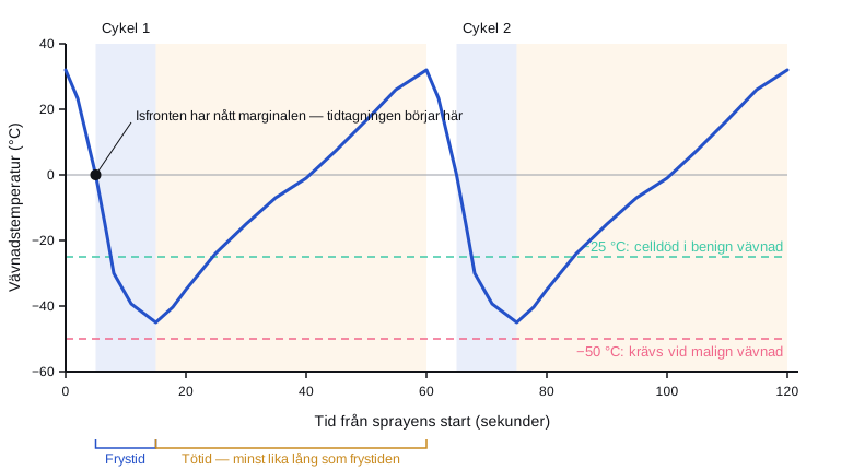
The freeze time is counted from the moment the ice front reaches the intended margin, not from the moment the spray starts. Let the tissue thaw completely between cycles — it is the freeze–thaw–freeze cycle that causes cell death, not the cold alone.
The temperature at the outer edge of the tissue is decisive: benign tissue requires about −25 °C for reliable cell death, malignant tissue about −50 °C. The outer limit of the ice front is only at 0 °C — the lethal temperature lies some way inside the visible ice front, and this is why the margin must be included in the freeze.
Contraindications
Absolute:
- Suspected melanoma or any other pigmented lesion of uncertain diagnosis.
- A lesion in which histology is needed for management.
- Cryoglobulinaemia, cold agglutinin disease, cold urticaria.
Relative:
- Basal cell carcinoma and squamous cell carcinoma — these can be treated with cryotherapy by an experienced practitioner in selected cases, but surgery is the standard.
- Dark skin: a risk of permanent hypopigmentation.
- Areas with poor circulation, above all the lower leg in a patient with peripheral arterial disease or diabetes — the wound may not heal.
- Over superficial nerves (the lateral aspects of the fingers, the ulnar groove, the neck of the fibula).
- The eyelid margins, the cartilage of the nasal tip, the free edge of the pinna.
- Autoimmune disease with Raynaud's phenomenon.
Preparation and equipment
- Liquid nitrogen in a flask (−196 °C) and a cryospray gun with interchangeable nozzles, or a cryoprobe for contact technique.
- Cotton buds for small lesions (these give a more superficial and less controllable depth of freeze).
- A size 15 scalpel or a pumice stone to pare down hyperkeratotic lesions.
- A cone or an otoscope speculum as a shield when treating near the eye or on an irregular surface.
- Petroleum jelly around the lesion to protect the surrounding skin.
- Eye protection and gloves for the operator.
Explain before treatment that it stings intensely during thawing, that a blister (sometimes a blood blister) is to be expected, that the area will be red and weeping for a week or so, and that hypopigmentation may be permanent. Document the consent.
Procedure
flowchart TD
  A[Skin lesion considered for cryotherapy] --> B{Pigmented or uncertain diagnosis?}
  B -- Yes --> C[Dermoscopy]
  C --> D{A confident benign diagnosis?}
  D -- No --> E[Biopsy or excision - do not freeze]
  D -- Yes --> F{Is histology needed for management?}
  B -- No --> F
  F -- Yes --> E
  F -- No --> G{A high-risk site such as the lower leg with vascular disease, a superficial nerve, an eyelid margin or cartilage?}
  G -- Yes --> H[Consider another method or referral]
  G -- No --> I{A hyperkeratotic surface?}
  I -- Yes --> J[Pare it down with a scalpel first]
  I -- No --> K[Freeze according to the table]
J --> K- Confirm the diagnosis. At the slightest uncertainty: dermoscopy, and if uncertainty persists a biopsy instead.
- Photograph the lesion — it will disappear, and the documentation is all that remains.
- Pare down any hyperkeratosis with a scalpel; nitrogen does not penetrate thick keratin.
- Protect the surroundings with petroleum jelly and, where necessary, a shielding cone.
- Hold the spray nozzle 1–2 cm from the skin, perpendicular to the surface.
- Spray intermittently until the ice front reaches the planned margin around the lesion. Start timing at that point.
- Allow complete thawing — the thaw time should be at least as long as the freeze time, usually 30–60 seconds.
- Repeat the cycle where indicated according to the table above.
- Check that the whole lesion, including the margin, has turned white.
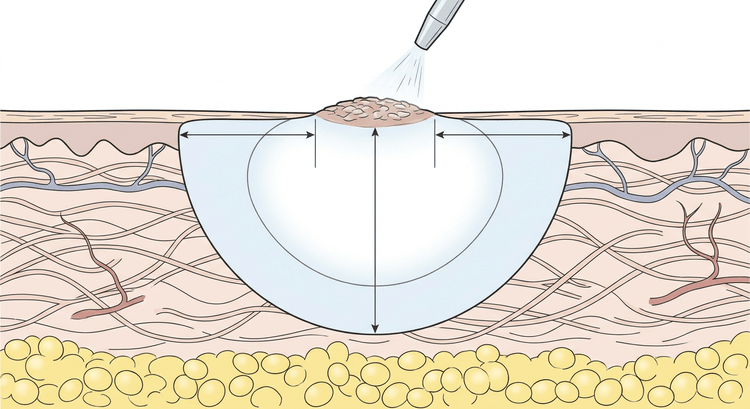
Contact technique with a cryoprobe gives a deeper and more controlled freeze on flat surfaces; spray technique is better on irregular surfaces and in the scalp.
Complications
| Complication | Frequency and comment |
|---|---|
| Pain during thawing | The rule rather than the exception |
| Blister, sometimes haemorrhagic | Expected; it must not be punctured routinely |
| Hypopigmentation | Common, often permanent — melanocytes are particularly sensitive to cold |
| Hyperpigmentation | Transient, commoner in people with darker skin |
| Scarring and atrophy | With too long a freeze time or too many cycles |
| Nerve injury with paraesthesiae | From freezing over superficial nerves; usually reversible within months |
| Infection | Uncommon |
| A wound that does not heal | Above all on the lower leg with impaired circulation |
| Recurrence | Common with too short a freeze time or too narrow a margin |
Aftercare and follow-up
Leave the blister in place as a biological dressing. If it is large and tense it can be drained with a sterile needle at its edge, but the roof must be left intact. Wash with soap and water, keep the area dry, and apply a dressing if the lesion rubs against clothing. Healing takes 1–2 weeks on the face and 3–6 weeks on the lower leg.
Arrange review after 4–8 weeks for actinic keratoses and where there is any question of tumour. A lesion that has not healed, or that recurs, must be biopsied, not frozen again. In widespread actinic damage, use field treatment with 5-fluorouracil, imiquimod or photodynamic therapy rather than spot cryotherapy.
Common pitfalls
- Freezing an undiagnosed pigmented lesion. A melanoma treated with cryotherapy loses both time and staging information.
- Too short a freeze time on hypertrophic lesions — the main cause of recurrence.
- Failure to pare down plantar warts makes the treatment ineffective.
- Counting the freeze time from the start of the spray instead of from the moment the ice front is achieved.
- Freezing too deeply on the lower leg in a patient with vascular disease — a cryotherapy wound can become a chronic leg ulcer.
- Freezing over superficial nerves without considering the anatomy.
- Failing to arrange review. An "actinic keratosis" that has not healed in eight weeks is a squamous cell carcinoma until proven otherwise.

