
What determines a lumbar puncture is rarely the needle itself. It is the decision whether to perform it now or after imaging, how the patient is positioned, which needle is chosen, and that the tubes go to the right place. In suspected bacterial meningitis it is the time to antibiotics that saves the patient — the puncture must never delay it.
Indications
- Suspected meningitis or encephalitis.
- Suspected subarachnoid haemorrhage with a normal or non-diagnostic CT.
- Suspected neuroborreliosis, neurosyphilis or another CNS infection.
- Suspected inflammatory disease: multiple sclerosis, Guillain–Barré syndrome, autoimmune encephalitis.
- Meningeal carcinomatosis and CNS involvement in haematological malignancy.
- Investigation of dementia with biomarkers (amyloid, tau, phospho-tau).
- Measurement of the opening pressure in suspected idiopathic intracranial hypertension.
- Therapeutic drainage in idiopathic intracranial hypertension and in assessment before shunt surgery.
Contraindications
- Clinical signs of herniation — deep unconsciousness with unreactive pupils, an altered breathing pattern, a rising blood pressure with bradycardia.
- A known or suspected intracranial space-occupying lesion with mass effect and midline shift.
- Ongoing anticoagulant treatment or marked coagulopathy. A platelet count below 50 × 10⁹/L is often quoted as the threshold, but the threshold varies between regions.
- Infection of the skin over the puncture site.
- Marked haemodynamic instability — stabilise the patient first.
CT before the puncture
Swedish practice differs here from several international guidelines. The Swedish clinical care programme for bacterial CNS infections states that CT must not be performed before lumbar puncture in suspected bacterial meningitis — not even with a reduced level of consciousness, focal neurological signs or seizures — because the delay costs more than the scan contributes. Only clinical signs of herniation halt the puncture, and antibiotics and a corticosteroid are then given immediately after blood cultures.
flowchart TD
  A[Suspected meningitis] --> B{Clinical signs of herniation?}
  B -- Yes --> C[Blood cultures, antibiotics and a corticosteroid immediately. No LP now]
  C --> D[CT, then LP once the patient's condition allows]
  B -- No --> E{Anticoagulant treatment or coagulopathy?}
  E -- Yes --> F[Correct it, or omit the LP. Treat on clinical suspicion]
  E -- No --> G[LP straight away, within 10-15 minutes of arrival]
  G --> H[Antibiotics immediately after the samples are taken; do not wait for the results]
For questions other than acute meningitis — an insidious onset, known malignancy, immunosuppression, papilloedema or new focal neurological signs — CT is performed first.
Preparation and equipment
- Informed consent, coagulation status and an up-to-date medication list.
- A sterile fenestrated drape, chlorhexidine in alcohol, sterile gloves, a face mask.
- An atraumatic needle (pencil point, Sprotte or Whitacre), 22–25 G. A cutting needle (Quincke) only when an atraumatic needle is unavailable or cannot be advanced.
- An introducer for the atraumatic needle — it is too soft to pass through the skin on its own.
- Local anaesthesia: lidocaine 10 mg/mL, a millilitre or two intracutaneously and along the needle track, with a fine needle.
- A manometer with a three-way tap when the opening pressure is to be measured.
- Four numbered sample tubes, and a light-protected tube if spectrophotometry is to be performed.
Procedure
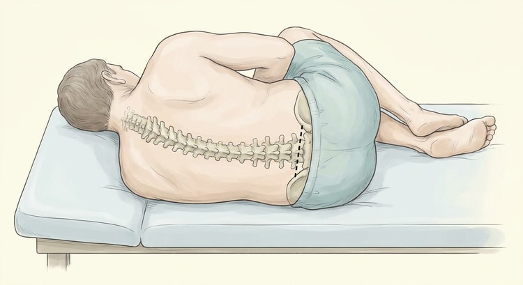
- Position the patient correctly. The lateral position with the back towards you, the knees drawn up, the chin on the chest, and the shoulders and hips stacked vertically. The back must be flexed like a bow — it is that flexion that opens the interspaces between the spinous processes. Sitting and leaning forward is an alternative when the lateral position does not work, but the opening pressure cannot be measured in the sitting position.
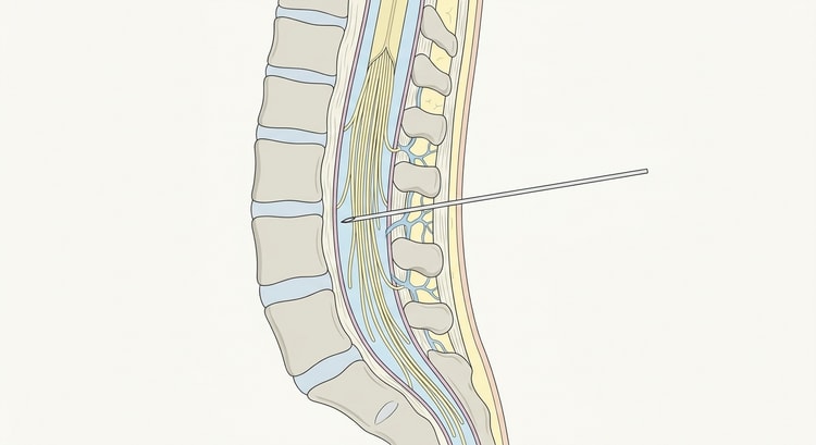
- Identify the level. Palpate both iliac crests and draw an imaginary line between them. The line crosses the midline at approximately L4. Puncture at the L3–L4 or L4–L5 interspace; in adults the spinal cord ends at T12–L1, so these levels encounter only the roots of the cauda equina. If in doubt: choose a level lower, never higher.
- Mark the puncture site, clean in wide circles and apply the drape.
- Anaesthetise the skin and the needle track. Wait a minute or so.
- Advance the needle in the midline with a slight cranial angulation, about 15 degrees towards the umbilicus. Use the introducer with the atraumatic needle. With a cutting needle the bevel must be turned parallel to the long axis of the body so that the dural fibres are separated rather than cut.
- Check the stylet every few millimetres as the resistance changes character. With an atraumatic needle there is no sharp give — instead, cerebrospinal fluid is the first sign.
- If you hit bone: withdraw the needle to the subcutaneous level without removing it completely, correct the angle and advance again. Never re-angle the needle while the tip is in the ligament.
- Measure the opening pressure if relevant, before any fluid is drained.
- Fill the tubes, withdraw the needle with the stylet in place, and apply a dressing.
Opening pressure
It is measured as soon as the cerebrospinal fluid appears, in the lateral position with the patient relaxed and the legs extended out of the curled-up position — sustained knee and hip flexion, crying and a Valsalva manoeuvre all falsely raise the pressure. Wait until the column is still and oscillating with respiration.
| Opening pressure | Interpretation |
|---|---|
| < 20 cm H₂O (< 200 mm H₂O) | Normal |
| 20–25 cm H₂O | A grey zone, not diagnostic |
| > 25 cm H₂O (> 250 mm H₂O) | Raised; required for the diagnosis of idiopathic intracranial hypertension |
Sample tubes and analyses
Number the tubes in the order in which they are filled. Procedures differ between laboratories — follow the local request instructions, but the principles are the same: chemistry and culture early, the cell count in a later tube, and one tube saved.
| Tube | Analysis | Comment |
|---|---|---|
| 1 | Albumin, glucose, lactate | With a simultaneous plasma glucose for the ratio |
| 2 | Culture and Gram stain, PCR | 1–2 mL; to the laboratory at once |
| 3 | Cell count with a polymorph/mononuclear differential | Analysed within 30 minutes; the cells break down |
| 4 | The saved tube, spectrophotometry, cytology, biomarkers | A cell count here as well distinguishes a traumatic tap from haemorrhage |
Where malignancy in the CNS is suspected, a considerably larger volume — at least 10 mL — is needed for the cytology to be meaningful.
Interpretation
| Finding | Normal | Bacterial meningitis | Viral meningitis | Tuberculous meningitis |
|---|---|---|---|---|
| Cell count (×10⁶/L) | < 5 | Often > 1,000 | 5–1,000 | 50–500 |
| Predominant cell type | — | Polymorphonuclear | Mononuclear | Mononuclear |
| Lactate (mmol/L) | < 3.5 | > 3.5, often higher | Normal or slightly raised | Raised |
| Albumin (g/L) | < 0.5 | Markedly raised, often > 1 | Normal or slightly raised | Raised |
| CSF/plasma glucose ratio | > 0.5 | < 0.4 | Normal | Low |
A normal cell count does not exclude bacterial meningitis in a patient who has presented very early or who is severely immunosuppressed. Antibiotics are given on clinical suspicion, not on the CSF result.
Subarachnoid haemorrhage
With a negative CT and a persisting suspicion, lumbar puncture is performed with spectrophotometry no earlier than 12 hours after the onset of symptoms — bilirubin forms in the cerebrospinal fluid only after that time, and an earlier sample may be falsely negative. The sample is protected from light and centrifuged promptly. The spectrophotometry distinguishes oxyhaemoglobin (an absorption peak at around 415 nm), which also arises from a traumatic tap in vitro, from bilirubin (around 455 nm), which requires time in living tissue and therefore indicates genuine haemorrhage. A decreasing admixture of blood between tube 1 and tube 4 is not on its own sufficient to exclude subarachnoid haemorrhage.
Complications
- Post-dural puncture headache — the commonest complication.
- Local back pain, usually transient.
- Transient radicular pain when the needle touches a nerve root.
- A traumatic tap that makes interpretation difficult.
- Epidural or spinal haematoma — very uncommon, but serious; suspect it in increasing back pain with neurological deficits.
- Infection, meningitis or an epidural abscess — rare with sterile technique.
- Herniation after a puncture despite raised intracranial pressure with mass effect.
Aftercare and follow-up
Bed rest is not needed. Lying down afterwards does not prevent post-dural puncture headache — this is well established and one of the most persistent myths about the procedure. Encourage a normal fluid intake and normal mobilisation.
Post-dural puncture headache typically appears within 1–2 days, is postural — worse when upright and relieved on lying down — and is often occipital or frontal with neck stiffness, nausea and sensitivity to sound. The risk is greatest in young women with a low BMI and lowest in people over 50. Management:
- Paracetamol and an NSAID, caffeine, plenty of fluid. Most symptoms resolve spontaneously within a week.
- For a severe or protracted headache: contact an anaesthetist for an epidural blood patch — the patient's own venous blood is injected epidurally at the level of the puncture and seals the dural hole. The effect often appears within hours and the treatment can be repeated.
- Reconsider the diagnosis if the headache is not postural, if there is fever, or if there are neurological deficits — venous sinus thrombosis, subdural haematoma and infection do occur after lumbar puncture.
Common pitfalls
- Waiting for a CT in suspected bacterial meningitis. The delay costs lives; the Swedish recommendation is to puncture immediately if there are no signs of herniation.
- Waiting for the CSF result before giving antibiotics.
- Poor positioning. Almost all failed punctures are due to insufficient flexion of the back or to the patient not lying perpendicular to the couch.
- The needle off the midline. If you hit bone immediately and superficially you are usually on a spinous process — palpate again rather than probing.
- An opening pressure measured sitting or with the legs drawn up — the value becomes falsely high and unusable.
- Spectrophotometry performed too early when subarachnoid haemorrhage is the question.
- Tubes left standing. The cell count is destroyed and the lactate changes when the sample stands.

