
Pericardiocentesis is performed on the basis of the haemodynamics, not the volume of fluid. A litre that has accumulated slowly can be asymptomatic, while 150 mL collected over an hour causes tamponade. The indication rests on the clinical picture together with echocardiographic signs of tamponade — not on how wide the rim of fluid looks. With ultrasound guidance, the point at which the fluid lies most superficially is chosen and the needle is followed all the way in, which has made the procedure considerably safer than the blind subxiphoid technique still described in older textbooks.
Indications
- Cardiac tamponade — the only absolute indication, and always urgent.
- A large pericardial effusion with symptoms despite medical treatment.
- Diagnostic drainage in suspected purulent pericarditis, tuberculosis or malignancy.
Tamponade is a clinical diagnosis supported by echocardiography:
| Finding | Comment |
|---|---|
| Hypotension, distended neck veins, muffled heart sounds | Beck's triad; complete in a minority |
| Tachycardia, tachypnoea, cold periphery | Almost always |
| Pulsus paradoxus above 10 mmHg | A fall in systolic blood pressure during inspiration; one of the most useful signs |
| Low voltage on the ECG, electrical alternans | Supportive; it does not exclude |
| Diastolic collapse of the right atrium and right ventricle | The cardinal echocardiographic finding |
| A dilated inferior vena cava without respiratory variation | Strongly suggests a raised filling pressure |
| Marked respiratory variation in mitral and tricuspid inflow | The echocardiographic counterpart of pulsus paradoxus |
flowchart TD
  A[Shock with a suspected pericardial effusion] --> B[Bedside echocardiography]
  B --> C{A pericardial effusion with signs of tamponade?}
  C -- No --> D[Look for another cause of shock]
  C -- Yes --> E{Suspected aortic dissection, myocardial rupture or trauma?}
  E -- Yes --> F[Emergency cardiothoracic surgery, not pericardiocentesis]
  E -- No --> G[Intravenous fluid, avoid intubation where possible]
  G --> H[Ultrasound-guided pericardiocentesis with a pigtail catheter]
  H --> I[Drain until the haemodynamics improve, leave the catheter in place]
Contraindications
In overt tamponade with shock there are no absolute contraindications other than the conditions in which the procedure is the wrong treatment:
- Aortic dissection with haemopericardium — drainage can increase the bleeding and worsen the prognosis. The patient must go to cardiothoracic surgery. In profound shock, very limited decompression may be considered in consultation with a cardiothoracic surgeon.
- Myocardial rupture after myocardial infarction, and traumatic haemopericardium — these require surgery; pericardiocentesis is at best a bridge to it.
- A small (under 10 mm), loculated or purely posterior effusion with no safe route for puncture — surgical drainage or pericardiotomy instead.
Relative: uncorrected coagulopathy, thrombocytopenia and ongoing anticoagulant treatment. In tamponade with shock the coagulation is corrected in parallel with the procedure, not before it.
Preparation and equipment
- Ultrasound with a sector probe (the same probe as for echocardiography) and a sterile probe cover.
- Continuous ECG, blood pressure and saturation monitoring; a defibrillator to hand.
- Sterile gown and gloves, chlorhexidine in alcohol 5 mg/mL, a large fenestrated drape.
- Lidocaine 10 mg/mL for local anaesthesia, 25 G and 21 G needles.
- An 18 G puncture needle, 7–9 cm (a spinal needle or the needle from a pericardiocentesis set), 10 mL and 20 mL syringes, a three-way tap.
- A Seldinger set with a guidewire, a dilator and a 5–8 F pigtail catheter, plus a drainage bag.
- Sample tubes for cell count, chemistry, culture and cytology, and blood culture bottles.
- Intravenous fluid and a vasopressor prepared.
The patient is positioned semi-recumbent, at 30–45 degrees — the fluid then collects anteriorly and inferiorly, which makes it easier to reach subxiphoid. Avoid sedation and positive pressure ventilation before decompression where possible: anaesthesia and intubation can precipitate circulatory collapse in a tamponaded patient, since positive airway pressure further reduces the venous return.
Procedure
Choice of access
The ultrasound, not tradition, determines the site. Scan subxiphoid, parasternally and apically, and choose the site where the rim of fluid is widest and lies most superficially, where the distance from the skin is shortest, and where no liver, lung or vessel lies in the path of the needle.
| Access | Puncture site | Direction | Comment |
|---|---|---|---|
| Subxiphoid | 1–2 cm below and to the left of the xiphoid process | 30 degrees to the abdominal wall, towards the left scapula | Avoids the pleura and the internal thoracic artery; the longest needle path, with a risk of injury to the liver and stomach |
| Apical | The 5th–6th intercostal space, 1–2 cm lateral to the cardiac apex | Towards the patient's right shoulder, close to the upper border of the rib | Often the shortest distance to the fluid; a risk of pneumothorax and of injury to the internal thoracic artery if one goes too medially |
| Parasternal | The 5th intercostal space, at least 3 cm lateral to the sternal edge | Perpendicular, inwards | A short path, but the internal thoracic artery runs 3–5 mm from the sternal edge |
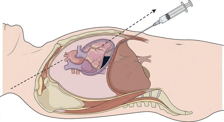
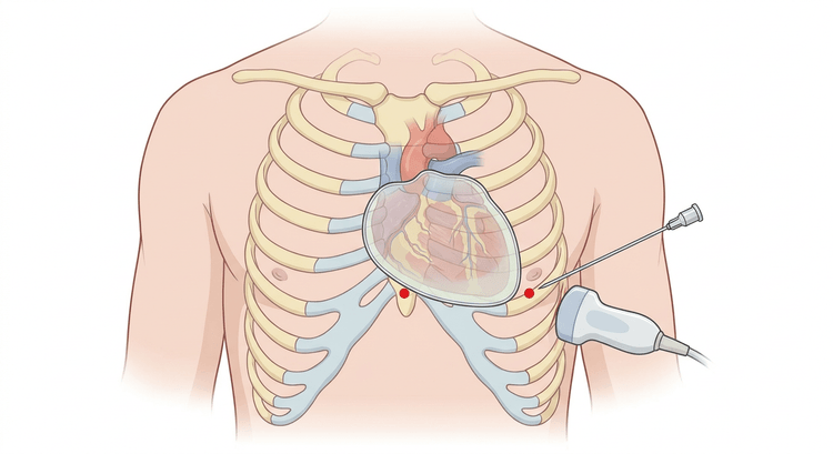
Ultrasound-guided puncture with the Seldinger technique
- Sterile preparation and draping. Confirm the fluid pocket again with a sterile probe and measure the distance from skin to fluid, noting the width of the rim of fluid in diastole.
- Anaesthetise the skin and the underlying tissue generously; aspirate throughout the advance of the anaesthetic needle.
- Advance the puncture needle along the planned path with continuous aspiration. With subxiphoid access, the needle is held flat, close to the undersurface of the sternum.
- Follow the needle tip on the screen the whole way. If the tip is not visible: stop, adjust the probe, and do not continue by feel.
- When fluid is aspirated, fix the needle with the hand resting against the patient and do not let go.
- Confirm the position before the guidewire is introduced. Inject 5–10 mL of agitated saline and see on the ultrasound that the bubbles appear in the pericardial space, not in the cardiac chambers. This is the single most important step in avoiding dilating into the right ventricle.
- Introduce the guidewire, withdraw the needle, dilate the tract and insert the pigtail catheter. Check the catheter position with ultrasound.
- Aspirate with a syringe and three-way tap, in aliquots. Even 20–50 mL often produces a dramatic haemodynamic improvement in tamponade — do not wait until everything is out before assessing the blood pressure.
- Connect the catheter to a drainage bag and secure it with a suture and a dressing.
- Record the ECG and blood pressure continuously, and note the volume drained and the appearance of the fluid.
ECG monitoring during the procedure
In the classical technique a chest lead was connected to the puncture needle and ST elevation was interpreted as epicardial contact. The method is unreliable and is replaced by ultrasound guidance, but continuous ECG monitoring is needed nonetheless: new ventricular ectopics or ventricular tachycardia during the advance mean that the needle is irritating the myocardium and must be withdrawn.
If the fluid is bloodstained
Pericardial fluid in haemopericardium does not as a rule clot, whereas blood aspirated from a cardiac chamber clots in the syringe. Let an aliquot stand for a few minutes. Compare the haematocrit with the patient's own as well. The saline injection under ultrasound is, however, the quickest and safest check.
Complications
| Complication | Comment |
|---|---|
| Puncture of a cardiac chamber or a coronary artery | The most serious complication; the risk falls markedly with ultrasound guidance |
| Pneumothorax, haemothorax | Above all with apical and parasternal access |
| Injury to the liver or stomach | From too steep an angle subxiphoid |
| Arrhythmia | Usually transient, on contact with the myocardium |
| Injury to the internal thoracic artery | With parasternal puncture closer than 3 cm to the sternal edge |
| Pericardial decompression syndrome | Acute pulmonary oedema and ventricular dysfunction after too rapid or too large a decompression |
| Infection, catheter-related pericarditis | Sterile technique; remove the catheter as soon as drainage has stopped |
Decompression syndrome is uncommon but serious. Do not drain more than about 1 litre at any one time, and decompress in aliquots with a haemodynamic assessment between each.
Aftercare and follow-up
- Leave the catheter in place and drain intermittently until the output falls below about 25–30 mL per day. An indwelling drain reduces the risk of recurrence.
- Echocardiography immediately after the procedure, and thereafter daily or on deterioration, to check that the fluid is not reaccumulating.
- A chest radiograph after apical or parasternal access to exclude a pneumothorax.
- Monitor on a unit with continuous ECG and blood pressure monitoring for the first 24 hours.
- Send the fluid for cell count and differential, protein and LDH, general culture including blood culture bottles, mycobacterial culture and PCR where suspected, and cytology in ample volume.
- Investigate the underlying cause: malignancy, tuberculosis, uraemia, bacterial infection, autoimmune disease, hypothyroidism, and the states following cardiac surgery or radiotherapy.
- A recurrent effusion despite adequate drainage is managed together with a cardiologist and a cardiothoracic surgeon: a pericardial window, balloon pericardiotomy or intrapericardial treatment.
Common pitfalls
- Waiting for imaging or transfer in a patient in tamponade shock. Bedside echocardiography and decompression on the spot save lives.
- Intubating and ventilating with positive pressure before decompression. This can precipitate cardiac arrest. Decompress first where possible.
- Pericardiocentesis in aortic dissection or myocardial rupture instead of emergency surgery.
- Blind subxiphoid puncture when ultrasound is available.
- Not confirming the needle position with agitated saline before dilatation — the commonest route to a pigtail catheter in the right ventricle.
- Too steep an angle subxiphoid, which directs the needle towards the liver and stomach instead of under the costal margin.
- Parasternal puncture too close to the sternal edge, injuring the internal thoracic artery.
- Draining too rapidly and too completely in a patient with a long-standing, large effusion.
- Removing the catheter immediately after drainage, and thereby missing the recurrence.


