
The insertion itself is rarely the difficult part — it is the confirmation of the position that determines whether the tube is safe. A misplaced tube in the airway produces no reliable symptoms in a patient with an impaired cough reflex, and the first feed then becomes a direct instillation into the lung. No tube may be used before its position has been verified by an approved method, and auscultation is not such a method.
Indications
- Decompression in ileus, subacute obstruction or gastric retention — the commonest indication on a surgical ward.
- Decompression in severe vomiting where the stomach does not empty.
- Intraoperatively and postoperatively after major abdominal surgery, according to local practice.
- Enteral nutrition and drug administration when the patient cannot swallow safely, for example after a stroke.
- Sampling of gastric contents and evacuation in poisoning (see the separate text on gastric lavage).
The tube is not, however, indicated routinely after uncomplicated abdominal surgery — it prolongs the time to return of bowel function without reducing the risk of complications.
Contraindications
| Type | Condition |
|---|---|
| Absolute | Suspected base-of-skull fracture or a marked midfacial fracture — the tube can pass through a fracture in the cribriform plate into the cranium |
| Absolute | Corrosive injury of the oesophagus or stomach after ingestion of a caustic substance |
| Absolute | Choanal atresia or an occluded nasal passage |
| Relative | Oesophageal varices or recent oesophageal or gastric surgery |
| Relative | Marked coagulopathy or ongoing epistaxis |
| Relative | Oesophageal stricture or a known diverticulum |
| Relative | Recent nasal or pharyngeal surgery |
Signs that should raise the suspicion of a base-of-skull fracture: periorbital bruising, retroauricular bruising, haemotympanum, CSF rhinorrhoea or CSF otorrhoea. Where this is suspected, the tube is passed through the mouth (orogastrically), never through the nose.
Preparation and equipment
- A tube of a suitable size. A wide-bore tube (Ch 14–18) of polyvinyl chloride for decompression and evacuation; a fine, soft tube (Ch 8–12) of polyurethane or silicone for nutrition, since it causes less mucosal irritation with longer-term use.
- A water-soluble lubricant, preferably with a local anaesthetic, and a decongestant nasal spray if the nasal passage is narrow.
- A 50–60 mL catheter syringe, a collection bag or suction, and a clamp.
- pH indicator paper with a scale that discriminates within the acidic range — ordinary litmus paper that only shows acid or alkaline is not sufficient.
- A fixation dressing for the nose, a vomit bowl, tissues, and a glass of water with a straw if the patient is awake and can swallow.
- Gloves, an apron, a visor. Suction must be out and working before you begin.
Inform the patient and agree a hand signal for a pause. Position the patient semi-recumbent with the chin slightly towards the chest — an extended neck directs the tube towards the larynx.
Procedure
- Inspect both nasal passages and choose the more patent one. Ask the patient about a previous nasal fracture or unilateral nasal obstruction.
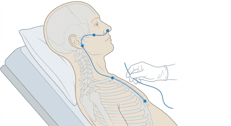
- Measure the depth of insertion by laying the tube from the tip of the nose to the earlobe and from there to the xiphoid process. Mark the distance on the tube. In adults the mark is typically around 50–60 cm.
- Lubricate the outermost 10–15 cm of the tube.
- Advance the tube perpendicular to the face, parallel to the floor of the nose — that is, straight backwards, not upwards towards the bridge of the nose. The resistance at the posterior pharyngeal wall after about 10–15 cm is expected.
- Ask the patient to flex the head forward and swallow repeatedly, preferably with sips of water if swallowing is safe. Advance the tube in time with the swallows.
- Stop immediately in the event of coughing, stridor, cyanosis or marked breathlessness — withdraw the tube to the pharynx and start again. If the tube emerges through the mouth it has coiled up in the pharynx; withdraw it and make a further attempt.
- Advance the tube to the marked level and secure it provisionally.
- Aspirate gastric contents and confirm the position (see below) before the tube is used and before it is secured definitively.
- Secure it against the ala of the nose without traction, so that the tube does not press against the edge of the nostril. Document the type and size of the tube, the nostril used, the depth of insertion in centimetres at the nostril, and the method by which the position was confirmed.
Confirming the position
The first-line method is pH measurement of the aspirate. Aspirate with a catheter syringe and drop the aspirate onto indicator paper.
- An acidic aspirate indicates a gastric position. The threshold differs between sources and regions: much of international practice accepts pH ≤ 5.5, while some national handbooks specify a stricter pH ≤ 4. Follow the threshold your own service has established.
- If no aspirate is obtained: turn the patient into the left lateral position, insufflate 10–20 mL of air and wait 15–20 minutes, or advance or withdraw the tube a few centimetres and try again.
- Proton pump inhibitors, H2 blockers and ongoing tube feeding all raise the pH and can give a falsely high value despite a correct position. A high pH therefore does not exclude a gastric position — but it is not sufficient as confirmation.
A radiograph is required when the pH cannot be assessed, when the patient has a reduced level of consciousness, impaired swallowing or an impaired cough reflex, when the tube is to lie in the small bowel, and before the tube is used for nutrition or drugs if the position has not been unequivocally confirmed by pH. The radiograph must be reviewed and documented by a doctor who comments specifically on the position of the tube tip — not as an incidental finding on an image taken for another reason.
Auscultation over the epigastrium after insufflation of air is not an approved means of confirming the position. The sound is transmitted to the epigastrium even when the tip lies in the oesophagus, in the bronchial tree or in the pleura, and the method has repeatedly preceded fatal misfeeding. It still appears in older procedure documents and is used in practice in many units — but it must not on its own form the basis for a decision to use the tube.
flowchart TD
  A[Tube advanced to the marked level] --> B{Is aspirate obtained?}
  B -- No --> C[Left lateral position, 10-20 mL of air, wait 15-20 min, adjust the depth]
  C --> B
  B -- Yes --> D{pH within the established acidic range?}
  D -- Yes --> E{Reduced consciousness, impaired swallowing or a small bowel position?}
  E -- No --> F[The tube may be used. Document the pH and the depth in cm]
  E -- Yes --> G[Radiographic confirmation before use]
  D -- No --> H{Treatment with a PPI or an H2 blocker, or ongoing tube feeding?}
  H -- Yes --> G
  H -- No --> G
  G --> I{Is the tube tip below the diaphragm in the stomach?}
  I -- Yes --> F
  I -- No --> J[Withdraw the tube and reposition it. Never use the tube]
Complications
- Malposition in the trachea or a bronchus with subsequent instillation of feed — the most serious and potentially fatal complication.
- Pneumothorax from perforation by a fine, stiff tube with a guidewire.
- Intracranial malposition in an undiagnosed base-of-skull fracture.
- Epistaxis, sinusitis and pressure necrosis of the ala of the nose from incorrect fixation.
- Oesophagitis, oesophageal stricture and mucosal ulceration with prolonged use.
- Aspiration during insertion or during feeding.
- Electrolyte disturbance and a hypochloraemic alkalosis with large aspirate losses that are not replaced.
- The tube coils up in the pharynx or comes out spontaneously, often in an anxious or confused patient.
Aftercare and follow-up
- Check the fixation and the external centimetre mark at least once per shift. A changed mark means that the position must be checked again.
- With nutrition: check the position before every new feeding session and after every episode of vomiting, vigorous coughing or pharyngeal suctioning.
- Flush the tube with water before and after every feed and every drug dose to prevent blockage. Do not crush modified-release tablets.
- Mouth care at least twice daily; the patient is often fasting and breathing through the mouth.
- Record the volume and appearance of the aspirate on the fluid balance chart when the tube is used for decompression. Replace larger losses as prescribed.
- The tube is removed once the underlying problem has resolved. In ileus: when the volume of aspirate has fallen, the patient has bowel sounds and is passing flatus, and the vomiting has stopped. Consider a trial period with the tube clamped before it is removed.
Common pitfalls
- Passing a nasal tube in a suspected base-of-skull fracture. Look for periorbital and retroauricular bruising and for CSF leakage before you start — choose the mouth at the slightest doubt.
- Auscultation as the only means of confirming position. A gurgling sound over the epigastrium proves nothing.
- Directing the tube upwards along the bridge of the nose instead of backwards along the floor of the nose — it hurts a great deal and gets nowhere.
- An extended neck during insertion, which opens the route towards the larynx.
- Not measuring the depth before insertion, thereby leaving the tip in the oesophagus, with reflux and aspiration as a consequence.
- Interpreting a high pH as malposition without taking PPI treatment into account — and conversely, interpreting it as acceptable without a radiograph.
- Not documenting the centimetre mark at the nostril. Without a baseline it is impossible to detect that the tube has slipped.
- Continuing to push despite coughing or a falling saturation. Withdraw and start again.

