
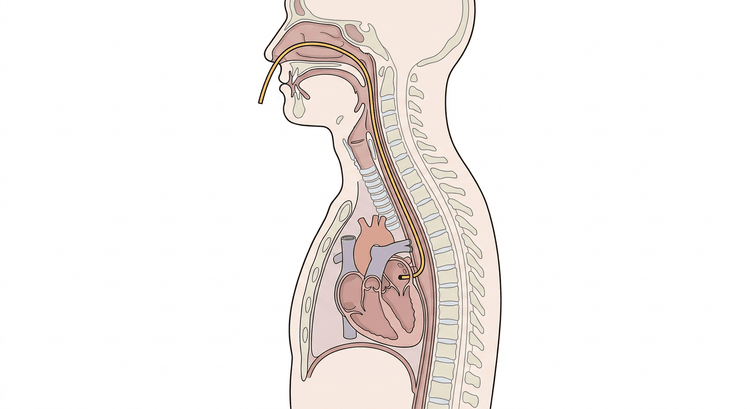
The oesophagus runs directly behind the left atrium. An electrode placed in the oesophagus at the right level therefore records atrial depolarisation with an amplitude the surface ECG can never achieve, and makes it visible even when it lies buried in the QRS complex or the T wave. The whole value of the investigation lies in the ratio between the number of atrial and ventricular complexes — more atrial than ventricular complexes, the same number, or fewer. That question determines the diagnosis, and it is asked of a patient who is haemodynamically stable enough for there to be time to ask it.
Indications
- Wide-complex tachycardia where ventricular tachycardia must be distinguished from supraventricular tachycardia with bundle branch block or aberrancy.
- Narrow-complex tachycardia where the P waves cannot be discerned on the surface ECG and the RP interval decides between AVNRT, orthodromic AVRT and atrial tachycardia.
- Suspected atrial flutter with a rapid, regular ventricular rhythm in which the flutter waves are concealed.
- A recurrent unclear wide- or narrow-complex tachycardia in which the mechanism needs to be documented before referral to an arrhythmia unit.
- Assessment of the effect of adenosine, which can be given during the recording and unmask the atrial activity.
The investigation is diagnostic, not therapeutic. An unstable patient should be cardioverted, not investigated.
Contraindications
- Haemodynamic instability — hypotension, shock, impaired consciousness or pulmonary oedema. Treat the arrhythmia first.
- Known stricture or tumour of the nose, pharynx or oesophagus.
- Oesophageal varices.
- Recent surgery of the nose, pharynx or oesophagus.
- Marked nausea, anxiety or communication difficulties that prevent the patient from cooperating.
- Suspected base-of-skull fracture or marked coagulopathy argues against the nasal route; the oral route may then be considered.
Preparation and equipment
- A 12-lead ECG machine able to print at 100 mm/s.
- A sterile oesophageal electrode, usually with colour-coded connectors (red and black) and a stiffening stylet.
- A Rostock filter or an equivalent amplifier unit between the electrode and the ECG machine.
- Lidocaine spray for the pharynx and lidocaine gel for the electrode.
- A glass of water with a straw, a vomit bowl, tissues, suction.
- Monitoring with ECG, blood pressure and saturation throughout the investigation.
Explain to the patient what is going to happen and that the most important thing is to swallow on command. A patient who understands the procedure tolerates it considerably better than one who is taken by surprise.
Procedure
- Connect the patient to the 12-lead ECG as usual and record a resting ECG of the arrhythmia before the electrode is inserted.
- Disconnect the V5 electrode from the chest and connect it to the "OUT" terminal of the filter. The two connectors of the oesophageal electrode are connected to the inputs of the filter ("IN−" and "IN+"). The oesophageal signal is thereby displayed in the V5 channel on the printout.
- Set the filter: a frequency range of about 15–40 Hz, and adjust the gain so that the atrial deflections are clear without clipping.
- Estimate the depth of insertion by measuring from the ala of the nose over the earlobe to the middle of the sternum. The depth is usually around 30–40 cm. Mark the measurement on the electrode.
- Anaesthetise the pharynx with lidocaine spray and lubricate the electrode generously with lidocaine gel.
- Introduce the electrode along the floor of the nose, parallel with the palate and straight backwards, not upwards towards the bridge of the nose.
- When the tip reaches the pharynx: let the patient take a mouthful of water and hold it. Advance the electrode at the very moment the patient swallows — the pharyngeal muscles then guide the tip into the oesophagus rather than into the trachea. Coughing, stridor or an inability to speak means malposition; withdraw and start again.
- Remove the stylet once the electrode is at approximately the intended depth.
- Adjust the position centimetre by centimetre, up and down, until the atrial deflections reach maximal amplitude. In the optimal position the atrial deflection is larger than the ventricular deflection in the oesophageal channel — that is the proof that the tip lies correctly.
- Print at 100 mm/s and for long enough to be able to count the complexes. Always compare the oesophageal channel with the simultaneously recorded surface leads on the same strip.
- If matters remain unclear, adenosine can be given intravenously during the recording; the transient AV block unmasks the atrial activity. Have a defibrillator available.
- Withdraw the electrode once the recording is complete and document the finding.
Interpretation
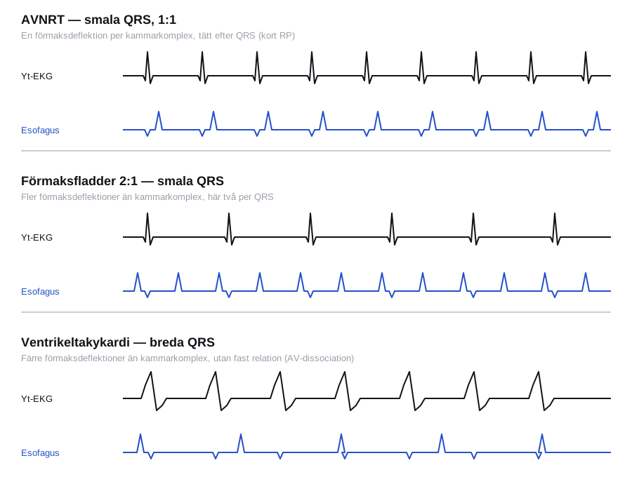
Always start from the same question: how many atrial deflections are there for each ventricular complex?
Wide QRS complexes
| Finding in the oesophageal channel | Interpretation |
|---|---|
| Fewer atrial than ventricular complexes, with no fixed relationship | AV dissociation — ventricular tachycardia |
| More atrial than ventricular complexes | Supraventricular tachycardia with bundle branch block or aberrancy, usually atrial flutter or atrial tachycardia with block |
| A 1:1 relationship | Diagnostically ambiguous: it may be VT with retrograde 1:1 conduction, SVT with aberrancy, or antidromic AVRT. Take the clinical picture, previous ECGs and the QRS morphology into account |
A 1:1 relationship does not exclude ventricular tachycardia. In a wide-complex tachycardia without definite AV dissociation the general rule still applies: treat as VT until the contrary is shown.
Narrow QRS complexes
| Finding | Interpretation |
|---|---|
| 1:1 with a short RP interval (RP shorter than PR, the atrial deflection closely after the QRS) | AVNRT, typically with a very short RP; orthodromic AVRT with a somewhat longer RP |
| 1:1 with a long RP interval (RP longer than PR) | Sinus tachycardia, ectopic atrial tachycardia or atypical AVNRT |
| More atrial than ventricular complexes, regular | Atrial flutter or atrial tachycardia with block. Count the atrial rate: around 250–300 per minute suggests typical flutter |
| Irregular, chaotic atrial deflections with no discernible baseline | Atrial fibrillation |
Always count the complexes on a sufficiently long strip. The fact that the degree of block alternates between 2:1 and 4:1 during the recording is in itself a strong argument for flutter.
Complications
Complications are uncommon. What the patient chiefly experiences is discomfort during insertion.
- Discomfort, retching and vomiting, with a risk of aspiration in a patient who cannot protect the airway.
- Epistaxis from the mucosa of the nasal passage.
- Malposition in the trachea with coughing and stridor — withdraw immediately.
- Hypersensitivity to lidocaine.
- Vasovagal reaction with bradycardia and a fall in blood pressure.
- Mucosal injury or, very rarely, perforation from forceful insertion or an undiagnosed stricture.
- That the investigation delays treatment in a patient who is deteriorating — the most serious risk, and the only one that is entirely avoidable.
Aftercare and follow-up
- Check the nose for bleeding and that the patient swallows and speaks normally.
- The patient can eat and drink as soon as the pharyngeal anaesthesia has worn off, usually after about an hour.
- Keep and scan the strip. An oesophageal ECG that shows the mechanism is often the only document available when the patient is later seen in the arrhythmia clinic, and it may determine whether ablation is offered.
- Document the atrial rate, the ventricular rate, the relationship between them, the RP interval in narrow-complex tachycardia, and the effect of adenosine if given.
- Treat the arrhythmia according to the mechanism established and consider referral for electrophysiological study.
Common pitfalls
- Performing the investigation in an unstable patient instead of cardioverting.
- Accepting the first position. If the electrode lies wrongly, the atrial deflection is small and the whole point is lost — move it until the deflection is maximal.
- Printing at 50 mm/s. At a high rate the atrial and ventricular complexes merge; 100 mm/s is needed to separate them.
- Looking at the oesophageal channel alone. Without simultaneously recorded surface leads it is impossible to decide what is atrial and what is ventricular.
- Interpreting a 1:1 relationship in a wide-complex tachycardia as proof of SVT. It is compatible with VT with retrograde conduction.
- Missing a varying degree of block because the strip is too short.
- Directing the electrode upwards towards the bridge of the nose instead of straight backwards along the floor of the nose — this hurts and causes epistaxis.
- Advancing the electrode when the patient is not swallowing, which increases the risk of malposition in the trachea.

