
The laryngeal mask is the single most important tool when intubation fails, since it restores oxygenation without requiring a view down into the larynx. The difficult airway rarely kills because the tube would not pass, but because the team keeps trying to intubate instead of changing the plan. The aim is always oxygenation, never intubation in itself — and every difficult airway algorithm is at bottom a timetable for when to stop doing what you are doing.
Indications
The laryngeal mask:
- A rescue airway when intubation has failed and bag-mask ventilation is difficult or inadequate.
- The primary airway in cardiac arrest when the person ventilating is not experienced in intubation.
- Planned anaesthesia for short procedures in a fasted patient at low risk of aspiration.
- A conduit for fibreoptically assisted intubation in a known difficult airway.
The difficult airway algorithm is activated in unexpectedly difficult laryngoscopy, difficult bag-mask ventilation or failed intubation — and already at the planning stage when the MACOCHA score or the LEMON assessment indicates that difficulties are likely.
Contraindications
The laryngeal mask is a compromise and does not protect the airway completely. Relative contraindications:
- A high risk of aspiration: a non-fasted patient, ileus, third-trimester pregnancy, marked reflux.
- A need for high airway pressures, for example severe ARDS, marked obesity or a stiff chest wall.
- Glottic or subglottic obstruction — the mask sits above the obstruction and solves nothing.
- A mouth opening below about 2 cm.
- Extensive pharyngeal pathology: tumour, abscess, marked swelling, previous radiotherapy.
In the "can't intubate, can't oxygenate" situation there are no contraindications — a laryngeal mask is inserted even with a full stomach.
Preparation and equipment
- A laryngeal mask of the correct size plus one size above and one below. Second-generation masks with a separate drainage channel (i-gel, LMA Supreme, LMA ProSeal) are the first choice in emergencies, since they allow the stomach to be decompressed and tolerate higher pressures.
- Lubricant on the dorsal surface of the mask, a syringe for the cuff in the models that have one, a cuff manometer.
- A videolaryngoscope with both a Macintosh-like and a hyperangulated blade, and a stylet shaped to match the hyperangulated blade.
- A bougie, tubes in several sizes, an oropharyngeal airway and a nasopharyngeal airway.
- Surgical airway equipment out and opened in a known or suspected difficult airway: a size 10 scalpel, a bougie and a cuffed size 6.0 tube.
- Capnography, suction, neuromuscular blockers, and a person designated to keep time and read out the number of attempts.
Laryngeal mask sizes
| Size | Patient weight | Maximum cuff volume (air-filled mask) |
|---|---|---|
| 1 | under 5 kg | 4 mL |
| 1.5 | 5–10 kg | 7 mL |
| 2 | 10–20 kg | 10 mL |
| 2.5 | 20–30 kg | 14 mL |
| 3 | 30–50 kg | 20 mL |
| 4 | 50–70 kg | 30 mL |
| 5 | 70–100 kg | 40 mL |
| 6 | over 100 kg | 50 mL |
The volumes are maxima — inflate to a seal and check that the cuff pressure does not exceed 60 cmH₂O. Gel-cuffed masks of the i-gel type have no cuff to inflate and are chosen on weight alone. When in doubt between two sizes in an adult: choose the larger, since a mask that is too small leaks and gives a poorer seal.
Procedure
Insertion of the laryngeal mask
- Check the mask, remove air from the cuff so that it is flat with a slightly upturned leading edge, and lubricate the dorsal surface.
- Position the head in the sniffing position if the neck may be moved.
- Ensure that the patient is sufficiently deeply sedated or relaxed — a half-awake patient coughs, clenches the jaw and develops laryngospasm.
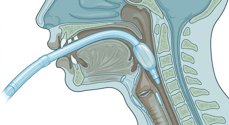
- Open the mouth, introduce the mask with the aperture facing the tongue and follow the hard palate with your index finger as a guide.
- Advance the mask downwards along the posterior pharyngeal wall in a smooth movement until a clear resistance is felt — the tip of the cuff then lies at the oesophageal inlet.
- Inflate the cuff to a seal, connect the capnograph and ventilate gently.
- Confirm: a sustained capnography trace, chest rise, symmetrical breath sounds and no audible leak at normal inspiratory pressure.
- Pass a gastric tube through the drainage channel of a second-generation mask and secure the mask with tape or a tie.
If the mask leaks or the patient is ventilated poorly: remove it, bag-mask ventilate, and reinsert — no more than three attempts, preferably with a different size. Pumping more air into the cuff usually makes the leak worse by pushing the mask out of position.
Videolaryngoscopy
- A hyperangulated blade requires a stylet shaped to the curvature of the blade; a straight stylet will not turn the corner even if the view is perfect.
- Introduce the blade in the midline under direct vision in the mouth, then move your gaze to the screen. Do not advance it too far — a view taken too close to the glottis makes passing the tube impossible.
- The best picture is not always the best position from which to pass the tube. Withdraw slightly and settle for a grade 2 view if the tube will then pass.
- Keep the lens clean; secretions and misting are the commonest reason why the videolaryngoscope does not help.
The difficult airway algorithm
Management follows the four-step DAS algorithm, which also underlies the recommendations of the Swedish Society of Anaesthesiology and Intensive Care Medicine (SFAI) for the difficult airway in Sweden. Each plan has a ceiling on the number of attempts, and the ceiling must be counted out loud by a designated person.
flowchart TD
  A[Plan A: bag-mask ventilation and intubation] --> B{Intubation successful?}
  B -- Yes --> C[Confirm with capnography, continue]
  B -- No, after at most 3 plus 1 attempts --> D[Declare failed intubation out loud]
  D --> E[Plan B: a second-generation laryngeal mask, at most 3 attempts]
  E --> F{Oxygenation restored?}
  F -- Yes --> G[Stop and choose the route: wake the patient, intubate through the mask, or a surgical airway under controlled conditions]
  F -- No --> H[Plan C: two-person bag-mask ventilation, an oropharyngeal airway and full relaxation]
  H --> I{Can the patient be oxygenated?}
  I -- Yes --> J[Wake the patient if the situation allows]
  I -- No --> K[Declare can't intubate, can't oxygenate]
  K --> L[Plan D: surgical airway with scalpel, bougie and a size 6.0 tube]
Surgical airway in "can't intubate, can't oxygenate"
CICO must be declared out loud in exactly those words so that the whole team knows which situation applies. Give 100 % oxygen, ensure full muscle relaxation — a tense larynx may itself be the cause — and go to the neck without further attempts at intubation.
- Extend the neck if possible and expose the front of the neck.
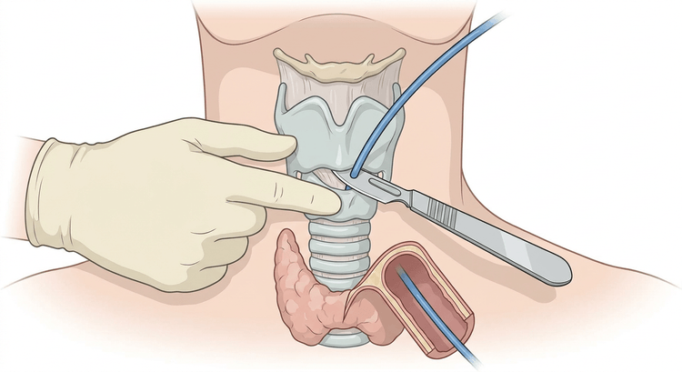
- Identify the membrane with the laryngeal handshake: grasp the larynx between the thumb and middle finger, stabilise it, and feel with the index finger for the depression between the lower border of the thyroid cartilage and the cricoid cartilage.
- A palpable membrane: stab the scalpel transversely through the membrane, rotate the blade 90° with the cutting edge downwards, and hold the scalpel in place as a guide.
- A non-palpable membrane (obesity, haematoma, swelling): make a vertical incision 8–10 cm long in the midline, separate the tissue bluntly with your fingers until the larynx can be palpated, and then continue as above.
- Pass the bougie along the scalpel blade a few centimetres down into the trachea — more than 10–15 cm increases the risk of injury.
- Railroad a cuffed size 6.0 tube over the bougie, inflate the cuff, connect the capnograph and ventilate.
- Confirm with a sustained capnography trace, secure the tube and summon ENT or cardiothoracic expertise for a definitive airway.
Complications
- Leakage and inadequate ventilation with the laryngeal mask, above all at high airway pressures.
- Aspiration of gastric contents — supraglottic devices protect incompletely.
- Sore throat, hoarseness and difficulty swallowing; usually transient.
- Injury to the lingual or hypoglossal nerve from an overinflated cuff.
- Laryngospasm and bronchospasm under too light anaesthesia.
- With a surgical airway: bleeding, malposition subcutaneously or in the mediastinum, injury to the larynx and cricoid cartilage, and later subglottic stenosis.
- Delay — the complication that causes hypoxic brain injury is almost always that the surgical airway was started too late.
Aftercare and follow-up
The laryngeal mask is removed either under deep anaesthesia or fully awake, never in the intermediate state in which laryngospasm is provoked. Suction the mouth clear before removal, not the pharynx behind the mask. After a surgical airway, intensive care is required, together with a decision about conversion to a tracheostomy or an endotracheal tube within the next 24 hours.
Always document a difficult airway: which technique worked and which did not, the laryngoscopic view, which adjuncts were used, and whether bag-mask ventilation was possible. The patient must be given written information and the airway must be recorded so that it is visible at the next episode of care. Report the event according to the local incident reporting procedure.
Common pitfalls
- Continuing with intubation attempts instead of moving on in the algorithm. Fixation on the tube is the central error in the difficult airway.
- Not declaring the situation out loud — "failed intubation" and "can't intubate, can't oxygenate" must be said in words, not implied.
- Too small a laryngeal mask with an overinflated cuff, which gives a worse seal rather than a better one.
- A laryngeal mask in a patient who is too lightly sedated.
- Giving up on bag-mask ventilation before an oropharyngeal airway, a two-handed grip and full relaxation have been tried.
- Searching for the cricothyroid membrane without stabilising the larynx with the laryngeal handshake.
- Too small a scalpel incision in the obese patient — here a vertical incision and blunt dissection are required, not a small hole in the skin.
- No documentation, no information to the patient, and therefore the same surprise next time.


