
What determines the outcome of an emergency intubation is rarely the laryngoscopy itself but what was done before it: how well the patient was preoxygenated, whether the difficulties were anticipated, whether the position and the equipment are in order, and whether there is an explicit plan for what happens when the first attempt fails. Desaturation and unrecognised oesophageal intubation are the two mechanisms that kill patients during airway management, and both are predictable.
Indications
- Inability to protect the airway: reduced level of consciousness, loss of pharyngeal reflexes, vomiting with a risk of aspiration.
- Inadequate oxygenation despite maximal oxygen or non-invasive ventilation.
- Inadequate ventilation with a rising pCO₂ and respiratory acidosis, or a markedly increased work of breathing with impending exhaustion.
- Threatened airway obstruction: inhalation injury, anaphylaxis, deep neck infection, an expanding haematoma, facial trauma.
- An anticipated course requiring a secured airway — transfer, heavy sedation, surgery — and cardiac arrest.
Contraindications
There are no absolute contraindications in the emergency situation. Consider alternatives when:
- Intubation is not consistent with the treatment plan — check any decision to limit treatment when time allows.
- There is complete upper airway obstruction where laryngoscopy will not succeed; plan for a surgical airway or awake fibreoptic intubation.
- A difficult airway is anticipated in a stable patient — awake fibreoptic intubation or anaesthetic back-up is then the right course, not RSI.
Assess the airway first
LEMON for a rapid bedside assessment:
| Letter | Assessment |
|---|---|
| Look | Facial trauma, beard, prominent incisors, a small chin, tumour, an irradiated neck |
| Evaluate 3-3-2 | 3 fingerbreadths of mouth opening, 3 fingerbreadths from chin to hyoid, 2 fingerbreadths from hyoid to the upper border of the thyroid cartilage |
| Mallampati | Class III–IV indicates increased risk |
| Obstruction/Obesity | Stridor, pharyngeal swelling, obesity |
| Neck mobility | Reduced neck mobility, a cervical collar, rheumatoid arthritis, ankylosing spondylitis |
MACOCHA is validated for the intensive care patient and totals 12 points: Mallampati III–IV (5 points), obstructive sleep apnoea syndrome (2 points), reduced neck mobility (1 point), mouth opening under 3 cm (1 point), coma (1 point), severe hypoxaemia below 80 % (1 point), and the intubator not being an anaesthetist (1 point). A score of 3 or more indicates a difficult intubation — call for help and get out the videolaryngoscope and plan B before you give the induction agent.
Preparation and equipment
Go through the equipment out loud with the team, not in your head.
- Functioning suction within reach under the pillow or by the right shoulder.
- A reservoir mask with a tight-fitting seal, a separate nasal cannula for apnoeic oxygenation, a bag with a PEEP valve, and an oropharyngeal airway.
- Laryngoscope: a videolaryngoscope as first choice where available, a direct laryngoscope with Macintosh blades 3 and 4 as back-up, always with the light checked.
- Tubes in three sizes, a stylet or bougie taken out and shaped before induction, a syringe for the cuff, a cuff manometer.
- A laryngeal mask of the correct size as a rescue airway, and surgical airway equipment (a size 10 scalpel, a bougie, a size 6.0 tube) out and visible.
- Capnography connected and calibrated.
- Working venous access, fluid and a vasopressor drawn up — push-dose noradrenaline or phenylephrine must be ready before induction, not looked for afterwards.
- Monitoring: ECG, pulse oximetry and blood pressure at short intervals.
Tube size and depth
| Patient | Internal diameter (mm) | Depth at the teeth (cm) |
|---|---|---|
| Adult woman | 7.0–7.5 | 21 |
| Adult man | 7.5–8.0 | 23 |
| Child, cuffed tube | age/4 + 3.5 | tube size × 3 |
| Child, uncuffed tube | age/4 + 4 | tube size × 3 |
| Term newborn | 3.0–3.5 | 6 + weight in kg |
Always have one size smaller to hand. The cuff is inflated to 20–30 cmH₂O measured with a manometer — a higher pressure causes mucosal ischaemia, a lower pressure leakage and aspiration.
Drugs for rapid sequence induction
The doses apply to an adult of normal weight and must be halved or reduced further in hypotension, hypovolaemia and in the frail elderly.
| Group | Agent | IV dose | Comment |
|---|---|---|---|
| Opioid | Fentanyl | 1–3 µg/kg | Blunts the sympathetic response; omitted in marked hypotension |
| Hypnotic | Propofol | 1.5–2.5 mg/kg | Marked fall in blood pressure; reduce to 0.5–1 mg/kg if unstable |
| Hypnotic | Ketamine | 1–2 mg/kg | First choice in hypotension, sepsis, asthma |
| Hypnotic | Thiopental | 3–5 mg/kg | Still used in some units |
| Neuromuscular blocker | Suxamethonium | 1–1.5 mg/kg | Onset 45–60 s, duration 5–10 min |
| Neuromuscular blocker | Rocuronium | 1.0–1.2 mg/kg | Onset about 60 s at the RSI dose, duration 45–70 min |
Suxamethonium is avoided in hyperkalaemia or suspected hyperkalaemia, in burns and denervation more than about 24 hours old, in prolonged immobilisation, spinal cord transection, muscular dystrophy and previous malignant hyperthermia. If rocuronium is chosen, sugammadex 16 mg/kg must be available for immediate reversal.
Procedure
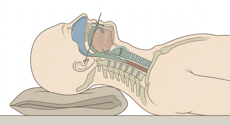
- Position the patient. The sniffing position in the patient of normal weight; a ramp in the obese patient so that the external auditory meatus reaches the level of the upper sternum. Raise the bed to your waist height and raise the head of the bed 20–30° if the circulation permits.
- Preoxygenate for at least 3 minutes with a tight-fitting mask and the highest possible flow, or 8 maximal breaths when time is short. If the saturation is inadequate: a PEEP valve or non-invasive ventilation during preoxygenation.
- Apply a nasal cannula at 15 L/min and leave it in place throughout the apnoeic phase — apnoeic oxygenation prolongs the time to desaturation.
- Allocate the roles out loud: who intubates, who gives the drugs, who manages suction and cricoid pressure, who keeps time and reads out the saturation.
- Give the induction agent and the neuromuscular blocker in rapid succession, flush afterwards, and wait for full relaxation before laryngoscopy. Inadequate relaxation is a common reason for a failed first attempt.
- Open the mouth, introduce the blade at the right corner of the mouth and sweep the tongue to the left. Follow the base of the tongue until the epiglottis is seen, place the tip of the Macintosh blade in the vallecula and lift along the long axis of the handle — never lever the blade against the upper incisors.
- Optimise the view with external laryngeal manipulation performed with your own right hand before the assistant takes over the grip.
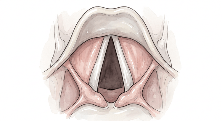
- Advance the tube under direct or video-assisted vision until the cuff has passed the vocal cords. In Cormack–Lehane grade 2–3: use a bougie, feel for the tracheal rings and railroad the tube over it.
- Inflate the cuff, connect the capnograph and ventilate.
- Confirm with capnography: a sustained waveform over at least six breaths is mandatory and takes precedence over auscultation, chest movement and misting of the tube. In cardiac arrest the values may be low, but the trace must have the right shape.
- Auscultate in the axillae and over the epigastrium, secure the tube, note the depth at the teeth and check the cuff pressure.
- Continue sedation, connect the ventilator and request a chest radiograph to check the tube depth.
Abandon the attempt and return to bag-mask ventilation if the saturation falls below 90 % or after about 30 seconds of laryngoscopy. No more than three attempts by the same person — after that something must change: a different operator, a different blade, a different position, or a switch to a laryngeal mask.
flowchart TD
  A[Tube inserted, cuff inflated] --> B{Sustained capnography trace over six breaths?}
  B -- Yes --> C[The tube is in the trachea]
  C --> D{Auscultation equal bilaterally?}
  D -- Yes --> E[Secure, check cuff pressure, chest radiograph]
  D -- No, silent on the left --> F[Withdraw the tube 1-2 cm, auscultate again]
  B -- No --> G[Assume oesophageal intubation]
  G --> H[Remove the tube, bag-mask ventilate with oxygen]
  H --> I{Can the patient be oxygenated?}
  I -- Yes --> J[New attempt with a changed strategy, call for help]
  I -- No --> K[Laryngeal mask, then the difficult airway algorithm]
Complications
- Unrecognised oesophageal intubation — the most serious and entirely avoidable complication.
- Hypoxaemia during the apnoeic phase, above all after inadequate preoxygenation.
- Hypotension and circulatory collapse after induction, particularly in hypovolaemia and after a high work of breathing before intubation.
- Aspiration of gastric contents.
- Endobronchial intubation, usually of the right main bronchus, with contralateral atelectasis.
- Injury to the teeth, lips and mucosa, laryngeal oedema, vocal cord injury and late tracheal stenosis.
- Arrhythmias, bradycardia in children, laryngospasm under light anaesthesia, and pressure necrosis of the trachea from an excessive cuff pressure.
Aftercare and follow-up
Continued sedation and analgesia according to the local scale, a lung-protective tidal volume of 6–8 mL/kg ideal body weight, the head of the bed raised 30°, and regular checks of the tube depth and the cuff pressure. A check blood gas after about 20 minutes and a chest radiograph to confirm that the tube lies above the carina.
Document the laryngoscopic view according to Cormack–Lehane, the number of attempts, which blade and which adjuncts were used, and whether bag-mask ventilation was possible. A patient who has been difficult to intubate must be given this in writing and it must be recorded in a way that will be seen at the next episode of care.
Common pitfalls
- Too short a period of preoxygenation — three minutes with a tight-fitting mask is not negotiable when time allows.
- Induction before plan B, the laryngeal mask and the surgical equipment are out.
- Relying on auscultation or misting of the tube instead of capnography.
- Continuing with repeated laryngoscopy attempts instead of oxygenating between attempts.
- Levering the blade against the incisors instead of lifting along the axis of the handle.
- Too small a dose of neuromuscular blocker, or laryngoscopy before the relaxation has taken effect.
- A full induction dose of propofol in a shocked patient.
- A forgotten nasal cannula during the apnoeic phase.
- No cuff manometer — the cuff is inflated by feel and is almost always overinflated.


