
Cerumen should be removed when it causes symptoms or obscures the tympanic membrane — not routinely. The wax has a protective function, and the commonest complication of ear syringing is that someone syringes an ear that did not need syringing. The second commonest is that the syringing is performed on a tympanic membrane that was already perforated.
Indications
Otoscopy: earache, hearing loss, ear discharge, dizziness, tinnitus, fever in a child without an obvious focus, and before every ear syringing.
Removal of cerumen:
- Hearing loss, a blocked sensation, itching, earache or dizziness attributable to a wax plug.
- The tympanic membrane cannot be inspected and the examination is needed for the diagnosis.
- Before audiometry or the fitting of a hearing aid.
- Recurrent otitis externa in which the wax retains moisture.
Contraindications
Absolute contraindications to irrigation:
- A known or suspected perforation of the tympanic membrane — including a history of grommets, ear discharge or pain on contact with water.
- A history of ear surgery in which the middle ear may be open (a mastoid cavity, mastoidectomy).
- An only hearing ear.
Relative (choose a manual technique or refer to ENT):
| Condition | Reason |
|---|---|
| Diabetes, immunosuppression | A risk of necrotising otitis externa |
| Anticoagulant treatment | Bleeding into the ear canal |
| Previous radiotherapy to the head and neck | Fragile skin, poor healing |
| Ear canal stenosis or exostoses | Narrow and hard to reach |
| Ongoing otitis externa | Pain, spread of infection |
| A small or uncooperative child | Movement causes injury |
Preparation and equipment
Otoscopy: an otoscope with a charged battery and clean disposable specula in several sizes. Use the largest speculum that will fit — it gives the most light and the best view.
Irrigation: a 30–60 mL syringe with a soft irrigation catheter, or an electric ear irrigator with pressure limitation, body-temperature water or saline (36–37 °C), a kidney dish, a towel, suction.
Manual technique: an operating otoscope or a head lamp with a loupe, a cerumen curette or loop, an angled hook, alligator forceps, a suction catheter (Frazier or Baron, 5 Fr).
Cerumenolytics (docusate sodium, sodium bicarbonate drops, olive oil, carbamide peroxide) are instilled 15–30 minutes before the procedure, or over a few days at home for a hard plug. Have the patient lie with the ear uppermost for 5 minutes after instillation.
Procedure
Otoscopy
- Inspect the outer ear, the tragus and the area behind the pinna before the otoscope is inserted — mastoid erythema and a protruding ear are not part of the tympanic membrane, but they are the most important finding when they are present.
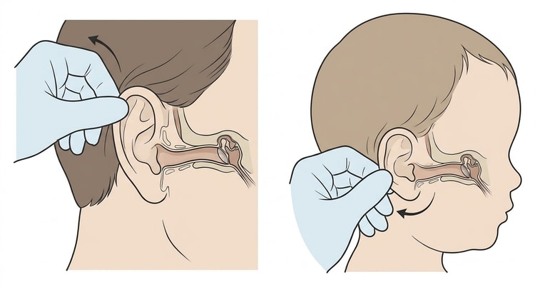
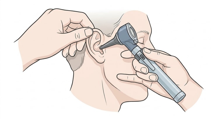
- Straighten the ear canal: pull the pinna upwards and backwards in adults, downwards and backwards in small children.
- Rest your hand against the patient's cheek so that the otoscope moves with the head if it moves.
- Insert the speculum under vision, to a maximum of about 8 mm.
- Follow a systematic order: the skin of the ear canal → the colour and translucency of the tympanic membrane → the light reflex → the handle and short process of the malleus → the pars flaccida → signs of fluid (a level, bubbles, retraction or bulging).
- Where needed, pneumatic otoscopy: a slight change of pressure with the bulb shows whether the tympanic membrane is mobile — an immobile membrane suggests fluid in the middle ear.
Irrigation
- Confirm that the tympanic membrane is intact if it can be seen at all, and ask specifically about a previous perforation or grommets.
- Instil a cerumenolytic and wait 15–30 minutes.
- The patient sits upright with a towel over the shoulder and holds the kidney dish against the neck below the ear.
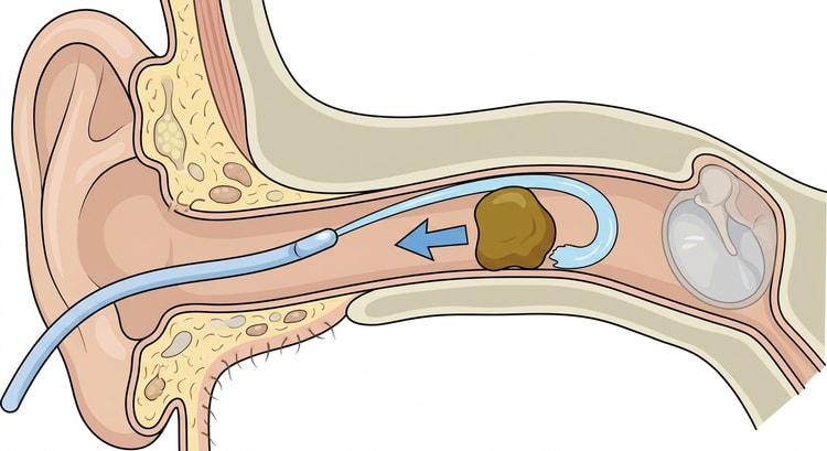
- Insert the catheter tip no more than 0.5 cm into the ear canal, not beyond the hair-bearing skin.
- Direct the jet upwards and backwards along the superior wall of the ear canal, past the plug — not straight at it. The water should get behind the wax and push it out.
- Use moderate pressure. Repeat in several rounds and inspect between each.
- Dry the ear canal and check the tympanic membrane. With an intact membrane, a few drops of isopropyl alcohol can speed the drying.
Manual removal
The work is more stable if the patient lies down with the head supported. Under direct vision: soft wax is removed with suction or a spoon-shaped curette, hard wax with a loop or an angled hook passed beyond the plug to draw it outwards. Never work with an instrument whose tip you cannot see.
| Consistency of the wax | Best technique |
|---|---|
| Soft, liquid | Suction |
| Firm but yielding | Curette or spoon |
| Hard, impacted | A cerumenolytic for a few days, then a loop or hook |
| Wax around a foreign body | No irrigation — see the separate guide |
Interpreting the tympanic membrane
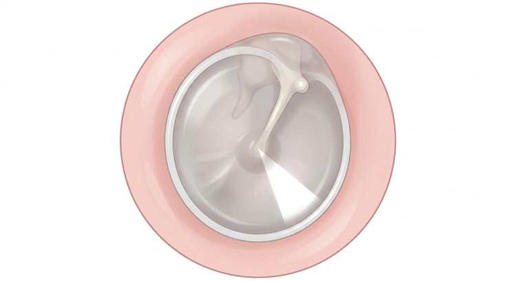
Always assess four things: colour, translucency, position (retracted, normal or bulging) and mobility. It is the combination that gives the diagnosis — redness alone is not sufficient, since a crying or febrile patient can have a red tympanic membrane without a middle ear infection.
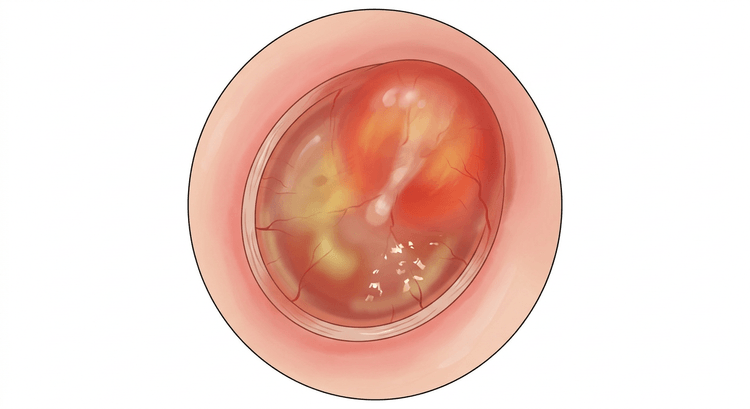
| Finding | Typical appearance | Interpretation |
|---|---|---|
| Normal | Pearly grey, translucent, light reflex forwards and downwards, mobile | No middle ear pathology |
| Acute otitis media | Opaque, red, bulging, immobile | The question of antibiotics according to the national recommendation |
| Otitis media with effusion | Dull, yellowish or amber, retracted, a fluid level or bubbles, reduced mobility | Fluid without acute infection — hearing check and follow-up |
| Perforation | A dark defect with a sharp edge, often in the pars tensa | Irrigation contraindicated |
| Grommets | A small plastic tube in the membrane, usually in the anterior inferior quadrant | Irrigation contraindicated |
| Myringitis | Blisters on the surface of the membrane | Painful, often viral |
| Cholesteatoma | A white flaking mass or a retraction pocket in the pars flaccida | Refer to ENT |
The tympanic membrane cannot be assessed at all if the wax obscures it. Describing a tympanic membrane you have not seen is a record entry that can become dangerous — write instead that it could not be inspected, and why.
Complications
- Perforation of the tympanic membrane — usually from too high an irrigation pressure or from instrumenting too deeply.
- A vestibular reaction with dizziness, nausea and bradycardia if the irrigation fluid is not at body temperature (caloric stimulation).
- Otitis externa, particularly if water remains behind residual wax.
- Necrotising otitis externa in patients with diabetes or immunosuppression — uncommon but serious.
- Bleeding and laceration of the ear canal, tinnitus, hearing loss.
Stop immediately in the event of severe pain, dizziness, tinnitus, sudden hearing loss or bleeding.
Aftercare and follow-up
Check the hearing after the procedure — preferably with the same method as before. Explain that cotton buds push the wax deeper and should be avoided. For recurrent wax plugs, olive oil 2–3 drops a week can be used prophylactically.
With a suspected perforation after irrigation: keep the ear dry, avoid drops containing neomycin, and review after 3–4 weeks. A persisting perforation or hearing loss is referred to ENT.
Common pitfalls
- Irrigating on an incomplete history. Ask explicitly about grommets, a previous perforation and ear surgery — otherwise you will find out afterwards.
- Cold or hot irrigation fluid provokes dizziness and nausea. Body temperature, every time.
- Instrumenting too deeply. Stay within 8 mm unless you have a full view.
- Neomycin-containing drops cause contact dermatitis in up to 13 % — choose another preparation.
- A water jet device or an oral irrigator as an irrigation tool — the pressure is sufficient to rupture the tympanic membrane.
- Continuing indefinitely. Decide in advance how many attempts are reasonable; a hard plug is better resolved by a few days of softening than by ten irrigations.
- Removing asymptomatic wax — without an indication there is only risk.

