
The single most important step in any eye injury is to measure and document the visual acuity before anything else is done. The second most important is to look for signs of perforation — Seidel's sign, an irregular pupil, a soft globe — because a perforated eye must never be instrumented, have its pressure measured, or be palpated.
Indications
- A sensation of something in the eye, watering, photophobia and redness after working with a grinder, a drill, a lawnmower, or in the wind.
- Suspected corneal abrasion.
- A red eye where the diagnosis is not obvious.
- Suspected acute glaucoma (tonometry).
Contraindications
Absolute contraindications to instrumental removal:
- Suspected penetrating injury or an intraocular foreign body — shield the eye with a rigid shield, give systemic antibiotics and contact an ophthalmologist urgently.
- A positive Seidel's sign.
- A foreign body centrally in the visual axis requiring an operating microscope.
Relative:
- A patient who cannot keep the gaze still.
- A deeply embedded body reaching the stroma or deeper.
- The absence of a slit lamp — the cornea should not be instrumented without magnification.
With a suspected chemical injury the priorities are reversed: irrigate first, for at least 30 minutes, and measure the pH in the conjunctival sac before anything else is done.
Preparation and equipment
- A visual acuity chart and a pinhole.
- A slit lamp with a cobalt blue filter; alternatively a magnifying loupe and a Wood's lamp.
- Oxybuprocaine or tetracaine drops (topical anaesthesia).
- Fluorescein strips.
- Sterile cotton buds.
- An eye spud or a 25–27 G needle on a 2 mL syringe as a handle.
- A low-speed rust ring burr if one is available.
- Sterile saline with a syringe for irrigation.
- An eye shield (never a pressure dressing when perforation is suspected).
- A tonometer: an applanation tonometer on the slit lamp, or a hand-held device (iCare, Tono-Pen).
Procedure
Systematic examination
- Visual acuity in each eye separately, with the patient's usual glasses and then through a pinhole. This is done before any drops.
- Inspect the eyelids, conjunctiva, cornea and anterior chamber. Note the pupil size, shape and reaction to light. A teardrop-shaped or peaked pupil suggests perforation. Look for hyphaema — blood with a fluid level in the anterior chamber, best seen with the patient sitting up.
- Ocular movements in all directions; pain on upward gaze may suggest an orbital floor fracture.
- Topical anaesthesia into the lower fornix.
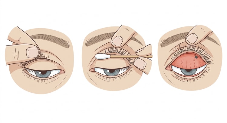
- Evert the upper eyelid. Ask the patient to look down. Grasp the eyelashes, press gently downwards on the upper border of the tarsal plate with a cotton bud, and at the same time fold the lid margin upwards and backwards towards the forehead. Double eversion is achieved by advancing the bud further downwards until the upper fornix is visible.
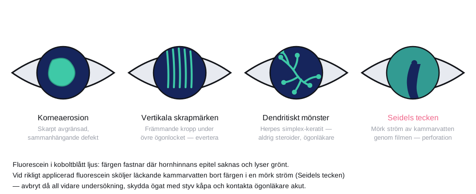
- Fluorescein and cobalt blue light. The dye adheres where the epithelium is absent. Vertical, linear scratch marks suggest a foreign body under the upper eyelid.
- Seidel's sign: apply fluorescein generously and look under blue light. A dark stream of aqueous humour breaking through the green film means perforation — stop everything else.
- Tonometry if acute glaucoma is suspected (normal 10–21 mmHg). Tonometry is contraindicated with suspected perforation or infection.
Removal
Proceed from the least to the most invasive technique.
- Irrigate with sterile saline — loose particles often come away.
- A moistened cotton bud with a rolling movement can lift off a superficial body from the conjunctiva or the cornea. Never use a cotton bud on an embedded corneal foreign body — it scrapes away a large area of epithelium.
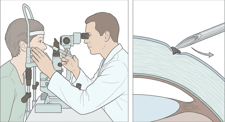
- An embedded body: the patient is positioned at the slit lamp with the forehead against the band and the chin in the rest. Support your working hand against the patient's zygoma or the bridge of the nose.
- Approach tangentially from the periphery, never through the visual axis, with a spud or a needle with the bevel upwards.
- Lift or scoop the body free with the tip. Work parallel to the surface of the cornea.
- A rust ring after an iron particle can be removed with a low-speed burr, but it does not have to be removed urgently — it can be dealt with by an ophthalmologist at the 24-hour review, and aggressive burring does more damage than the rust does.
- Check the visual acuity and Seidel's sign after the procedure.
Complications
- Corneal abrasion, often larger than the original injury.
- Corneal scarring with permanent visual impairment if the injury is central.
- Infectious keratitis and, rarely, endophthalmitis.
- Perforation from instrumenting too deeply.
- A retained rust ring with persistent inflammation.
- A missed intraocular foreign body — the most serious, and the only one that is entirely avoidable through the history (a high-velocity injury, metal on metal) and radiography or CT where suspected.
When to contact an ophthalmologist
Contact an ophthalmologist urgently, the same day, for any of the following:
- Suspected penetrating injury or an intraocular foreign body — a positive Seidel's sign, a teardrop-shaped or peaked pupil, a soft globe, prolapsed dark tissue in the wound edge, or a history of metal on metal, a grinder, an angle grinder or a hammer. Do not instrument, do not measure the pressure, do not palpate; apply a rigid shield, keep the patient fasting, give systemic antibiotics and arrange transfer.
- Hyphaema — blood in the anterior chamber after blunt trauma. A semi-upright position, no aspirin or NSAIDs, and ophthalmological assessment the same day; the risks are rebleeding and a rise in pressure.
- Visual loss not explained by a refractive error and not improved by a pinhole.
- A retained rust ring after an iron particle, or a foreign body that could not be removed.
- Dendritic fluorescein uptake or a corneal infiltrate, particularly in a contact lens wearer.
- A central injury in the visual axis, a chemical injury, or a persisting epithelial defect at the 24-hour review.
flowchart TD
  A[Eye injury or a red eye after trauma] --> B{Chemical injury?}
  B -- Yes --> C[Irrigate for at least 30 minutes, measure the pH in the conjunctival sac, contact an ophthalmologist]
  B -- No --> D[Measure and document the visual acuity in each eye separately]
  D --> E{Signs of perforation: Seidel's sign, a teardrop-shaped pupil,<br/>a soft globe, prolapsed tissue, a deep laceration track?}
  E -- Yes --> F[Do not instrument, do not measure the pressure, do not palpate.<br/>Rigid eye shield, keep fasting, systemic antibiotics,<br/>tetanus cover and urgent transfer to an eye unit]
  E -- No --> G{High-velocity injury, metal on metal,<br/>a grinder or a hammer?}
  G -- Yes --> H[Suspect an intraocular foreign body:<br/>CT of the orbit and ophthalmological assessment the same day]
  G -- No --> I{Hyphaema, visual loss, dendritic<br/>fluorescein uptake or a central injury?}
  I -- Yes --> J[Ophthalmological assessment the same day]
  I -- No --> K{A superficial foreign body that could be removed?}
  K -- Yes --> L[Antibiotic ointment and review,<br/>any residual rust ring assessed within 24 hours]
  K -- No, it remains or is deeply embedded --> J
Aftercare and follow-up
| Measure | Comment |
|---|---|
| Topical antibiotic | Chloramphenicol ointment or fluoroquinolone drops 3–4 times daily for 5–7 days. In contact lens wearers, a fluoroquinolone is chosen to cover Pseudomonas |
| Analgesia | Oral paracetamol and an NSAID |
| Cycloplegic | For marked photophobia and ciliary spasm, for example cyclopentolate |
| Dressing | Not needed, and it does not improve healing |
| Contact lenses | A break until the epithelium has healed and treatment has been completed |
| Follow-up | Review within 24 hours by an ophthalmologist if there is a persisting defect, a rust ring or a central injury |
Never prescribe topical anaesthetics for use at home — they delay healing and can cause corneal melting. Topical steroids must not be given without an ophthalmological assessment.
Give the patient clear instructions to seek urgent help if the pain increases, the vision deteriorates, the redness increases, or there is purulent discharge. Check the tetanus vaccination status.
Common pitfalls
- The visual acuity not measured before treatment — indefensible both legally and clinically.
- The upper eyelid not everted. Vertical scratch marks on the cornea without a visible foreign body mean that it is sitting under the lid.
- Seidel's sign not checked before instrumentation.
- A cotton bud on an embedded corneal foreign body.
- The history of a high-velocity injury overlooked — metal on metal requires CT of the orbit.
- Tonometry on a perforated eye.
- Anaesthetic drops sent home with the patient, or steroid drops without ophthalmological input.
- A chemical injury examined before irrigation. Irrigate first, examine afterwards.

