
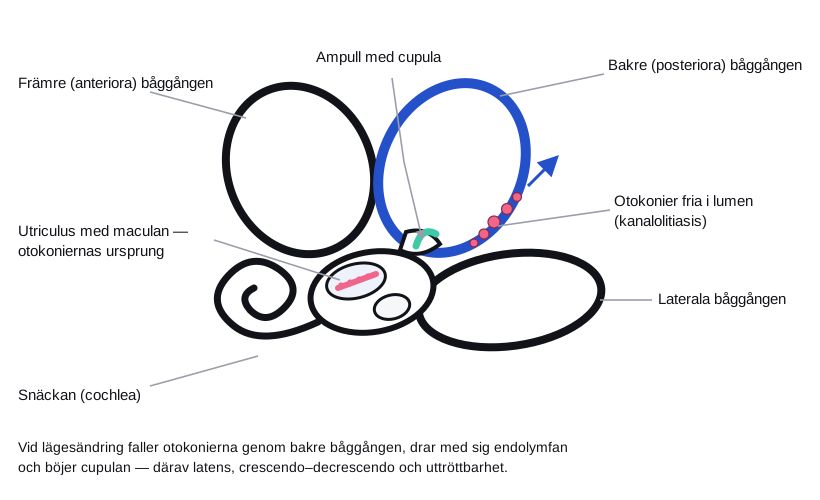
Benign paroxysmal positional vertigo is the commonest cause of vertigo in primary care and one of the few diagnoses that can be cured on the spot, in three minutes, without drugs. The diagnosis rests on the history, a positive Dix–Hallpike test and the absence of other neurological findings. The treatment is a repositioning manoeuvre.
Indications
- Brief attacks of vertigo, seconds to a minute or so, that are provoked by a change of position — turning over in bed, lying down, standing up, tilting the head backwards.
- A symptom-free interval between attacks, with possible residual unsteadiness.
- No hearing loss, tinnitus or neurological deficits.
In horizontal positional vertigo (lateral semicircular canal) the supine roll test is used for diagnosis, and the barbecue roll or the Gufoni manoeuvre for treatment.
Contraindications
Absolute:
- Unstable cervical spine injury, recent cervical spine surgery, atlantoaxial instability, severe rheumatoid arthritis of the neck.
- Symptomatic carotid stenosis, recent stroke or TIA in the vertebrobasilar territory.
- Retinal detachment.
Relative:
- Marked cervical restriction of movement or pain — use the Semont manoeuvre or the side-lying variant of the Dix–Hallpike test.
- Marked obesity or spinal disease that makes positioning impossible.
- Severe angina, heart failure or recent myocardial infarction.
Red flags — investigate further instead
| Finding | Suggests |
|---|---|
| Nystagmus that is purely vertical, downbeating or direction-changing | Central cause |
| Nystagmus without latency that does not fatigue | Central cause |
| New headache, neck pain, diplopia, dysarthria, dysphagia, ataxia | Posterior circulation stroke |
| Unilateral hearing loss or tinnitus | Ménière's disease, vestibular schwannoma, labyrinthitis |
| Continuous vertigo that does not come in attacks | Vestibular neuritis, central cause |
| Marked imbalance that makes walking impossible | Cerebellar infarction |
HINTS in continuous vertigo
HINTS is not a test for BPPV. It is used in the acute vestibular syndrome — continuous vertigo with spontaneous nystagmus, nausea and difficulty walking that has lasted hours to days — to distinguish vestibular neuritis from posterior circulation infarction. The test presupposes that the patient has spontaneous nystagmus at the time of examination and that the examiner is experienced with the head impulse test; performed on the wrong patient or by an inexperienced examiner it is unreliable. A single central feature is enough to proceed with urgent investigation, and a normal CT of the brain does not exclude an infarction in the posterior fossa.
flowchart TD
  A[Vertigo] --> B{Attacks provoked by a change of position<br/>with symptom-free intervals?}
  B -- Yes --> C[Dix-Hallpike test]
  C -- Upbeating torsional nystagmus with latency --> D[BPPV: treat with the Epley manoeuvre]
  C -- Atypical or no nystagmus --> E[Consider the lateral canal: supine roll test]
  B -- No, continuous vertigo with spontaneous nystagmus --> F[Acute vestibular syndrome: perform HINTS]
  F --> G{Head impulse test}
  G -- Abnormal with a corrective saccade --> H{Nystagmus and skew deviation}
  G -- Normal --> I[Central until proven otherwise]
  H -- Direction-fixed horizontal nystagmus, no skew --> J[Peripheral: vestibular neuritis]
  H -- Direction-changing or vertical nystagmus, or skew --> I
  I --> K[Urgent MRI with diffusion, contact the on-call stroke physician]
  J --> L[Symptomatic treatment and early mobilisation]
Preparation and equipment
No special equipment is needed. What is useful in practice:
- An examination couch that can be approached from all sides and on which the patient's head can hang over the end.
- A pillow to place under the shoulders as an alternative to a hanging head.
- Frenzel goggles or video-Frenzel if available — small beats of nystagmus can otherwise be hard to see, particularly when the patient fixates.
- A vomit bowl; nausea is common.
Tell the patient that the test will in all probability provoke intense but short-lived vertigo, and that this is precisely the point. Ask the patient to keep the eyes open and look straight ahead — without open eyes there is no diagnosis.
Procedure
The Dix–Hallpike test
- The patient sits upright on the couch, far enough forward that the head will extend beyond the end of the couch when laid down.
- Turn the head 45° to the right.
- Support the head and lay the patient quickly into the supine position with the head extended approximately 20–30° below the horizontal, maintaining the 45° rotation.
- Observe the eyes for 30–60 seconds.
- Sit the patient up again and observe the eyes for a further period — reversal of the nystagmus is common.
- Repeat on the left side.
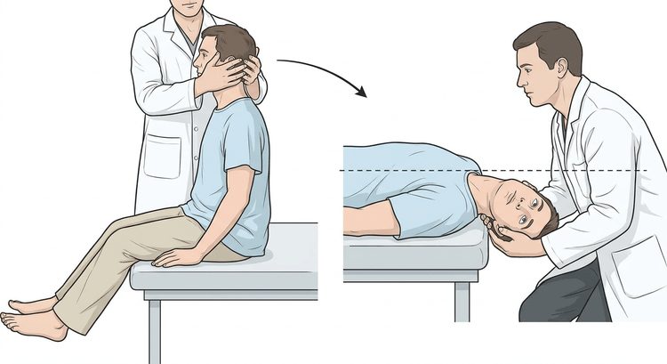
A positive test in the posterior canal is characterised by:
- A latency of 1–5 seconds before the nystagmus begins.
- Upbeating and rotatory (torsional) nystagmus with the fast phase beating towards the undermost, affected ear.
- A crescendo–decrescendo course that subsides within 30–60 seconds.
- Fatigability on repetition.
- Simultaneous subjective rotatory vertigo.
The ear that is downmost when the nystagmus is provoked is the affected one.
The Epley manoeuvre
Start from the side that was positive. Hold each position until the nystagmus and the vertigo have subsided, but at least 30 seconds, preferably 60.
| Step | Position |
|---|---|
| 1 | Sitting, head turned 45° towards the affected side |
| 2 | Quickly down into the supine position with the head extended 20–30°, the rotation maintained (= the Dix–Hallpike position) |
| 3 | Turn the head slowly 90° in the opposite direction, so that it lies 45° towards the unaffected side, still extended |
| 4 | Roll the patient onto the side towards the unaffected side and turn the head a further 90°, so that the nose points obliquely down towards the floor |
| 5 | Sit the patient slowly up with the chin slightly tucked down |
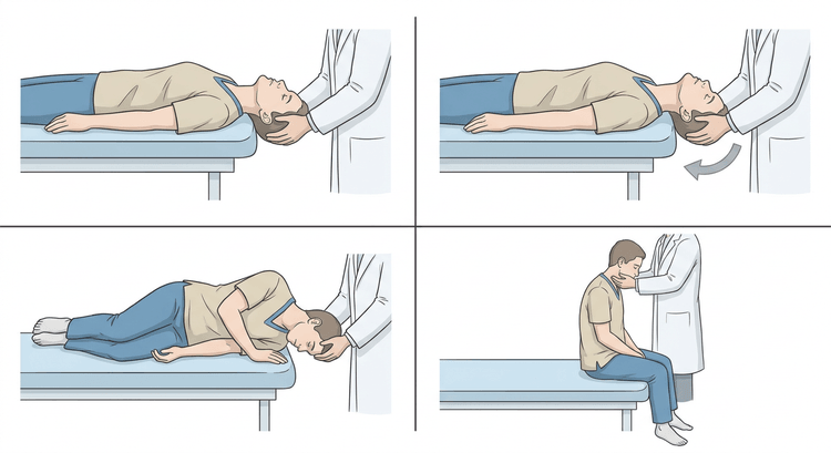
Nystagmus in the same direction as in the starting position during the manoeuvre indicates that the otoconia are moving the right way. Nystagmus in the opposite direction suggests that they are travelling back — start again.
Repeat the manoeuvre 1–3 times at the same visit, or until the Dix–Hallpike test is negative. The effect is immediate in the majority — around 80 % become free of symptoms after one or two rounds of treatment.
The Semont manoeuvre is an equivalent alternative, particularly when the neck does not permit extension: the patient sits on the edge of the couch with the head turned 45° away from the affected side, is laid rapidly onto the affected side and held there, and is then swung in one rapid movement across to the opposite side with the head rotation maintained.
Complications
- Intense vertigo, nausea and vomiting during the manoeuvre — the commonest.
- Conversion to the lateral canal (canal switch), 5–10 %: the vertigo changes character and becomes horizontal. It is diagnosed with the supine roll test and treated with the barbecue roll.
- Neck pain.
- Vasovagal reaction.
- Very rarely: a vascular event in the posterior circulation in a patient with vascular disease.
Pretreatment with an antiemetic (for example meclozine) may be considered in marked nausea, but sedatives and antihistamines must not be given routinely — they prolong central compensation and mask the diagnosis.
Aftercare and follow-up
The old restrictions about sleeping upright for two days or avoiding bending the head have not been shown to improve the outcome and are unnecessary. Instead, explain that a slight unsteadiness may persist for a few days and that it is improved by movement, not by rest.
Teach Brandt–Daroff exercises for home use in persistent or recurrent symptoms: sitting on the edge of the bed, quickly down onto the side with the head turned 45° upwards, 30 seconds, up, 30 seconds, down on the other side. Five repetitions, three times daily.
Arrange review after 1–2 weeks if symptoms persist. Recurrence affects around 15–30 % within a year and is treated in the same way. Refer to ENT or neurology in:
- Failure to respond after 2–3 rounds of treatment.
- Atypical nystagmus or any of the red flags above.
- Frequent recurrences.
Investigate for vitamin D deficiency in recurrent BPPV — supplementation reduces the recurrence rate.
Common pitfalls
- The patient closes the eyes during the test. Without visible eyes there is no diagnosis.
- Lowering the patient too slowly provokes no nystagmus and gives a falsely negative test.
- Too short an observation period. The latency may be up to five seconds, and that feels like a long time.
- Treating the wrong side after incorrect lateralisation — the affected ear is the one that lies downmost.
- Central nystagmus interpreted as BPPV. Purely vertical, direction-changing or non-fatiguing nystagmus should be investigated, not manoeuvred.
- Betahistine and sedatives instead of a manoeuvre. Drugs have no place in the treatment of BPPV.
- Positions held too briefly during the Epley manoeuvre — 30 seconds is a minimum, not a recommendation.

