
Spirometry is the only investigation that distinguishes obstructive from restrictive impairment of lung function, and COPD cannot be diagnosed without it. The quality of the test determines its entire value: a poorly performed manoeuvre gives a falsely low FVC and thus a falsely normal FEV₁/FVC. Set aside time, coach actively, and do not accept a trace you would not be prepared to show a colleague.
Indications
- Investigation of chronic cough, dyspnoea, wheeze or reduced exercise capacity.
- Suspected asthma or COPD — diagnosis and follow-up.
- Risk groups: current or former smokers over 40 with respiratory symptoms, and occupational exposure to dust, gas or fumes.
- Preoperative assessment before thoracic or upper abdominal surgery in a patient with known lung disease.
- Follow-up of treatment effect and of disease progression.
- Assessment of pulmonary involvement in systemic disease, for example rheumatoid arthritis or scleroderma, and during treatment with pulmonary toxic drugs.
Contraindications
Forced expiration raises the pressure in the thorax, the abdomen, the skull and the eyes. Postpone the test in:
| Condition | Recommended delay |
|---|---|
| Myocardial infarction or unstable angina | 1 month |
| Pneumothorax | 1 month after resolution |
| Pulmonary embolism, until anticoagulated and stable | An individual assessment |
| Eye, ear, abdominal or thoracic surgery | 1 month |
| Neurosurgery or an intracranial aneurysm | Contact the responsible specialist |
| An aortic aneurysm > 6 cm | Relative — avoid forced manoeuvres |
| An ongoing respiratory infection with a risk of transmission | Until symptom-free |
| Haemoptysis of unclear cause | Until investigated |
Relative obstacles also include dementia, marked hearing loss and an inability to understand the instructions — spirometry requires active cooperation.
Preparation and equipment
Equipment: a calibrated spirometer with a daily volume check (a 3-litre calibration syringe), a disposable mouthpiece with a bacterial filter, a nose clip, a chair with armrests, a bronchodilator with a spacer, and current reference values (GLI 2012 is the standard).
Patient preparation — given with the appointment letter:
| Drug or habit | Period to be withheld before the test |
|---|---|
| Short-acting beta-2 agonist (salbutamol, terbutaline) | 4–6 hours |
| Short-acting antimuscarinic (ipratropium) | 12 hours |
| Long-acting beta-2 agonist (formoterol, salmeterol) | 24 hours |
| Ultra-long-acting beta-2 agonist (indacaterol) | 36 hours |
| Long-acting antimuscarinic (tiotropium, umeclidinium, glycopyrronium) | 36–48 hours |
| Smoking | 1 hour |
| Vigorous physical exertion | 30 minutes |
| A large meal | 2 hours |
Withholding applies only to diagnostic spirometry with a reversibility test. At follow-up during ongoing treatment, the test is performed on the usual medication — note this in the report.
Record the height without shoes, the weight, the age, the sex and the ancestry before the measurement; the reference values are based on them. In kyphosis, or when the patient cannot stand, the arm span divided by 1.06 is used as the height.
Procedure
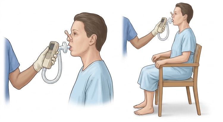
- Explain and demonstrate. Show for yourself what a maximal blow looks like. The patient sits upright with both feet on the floor and must not lean forward during expiration.
- Apply the nose clip and have the patient seal the lips tightly around the mouthpiece without the tongue blocking it.
- Quiet breathing for a few breaths until the tidal volume is stable.
- A maximal inspiration to total lung capacity — rapid, with a minimal pause at the top (< 1 second).
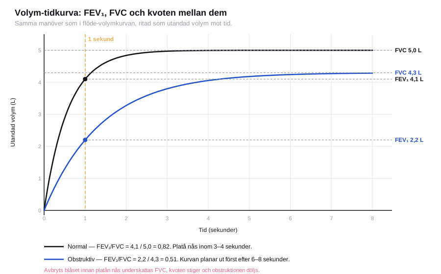
- A maximal forced expiration: an explosive start and then continued blowing with loud coaching until a plateau is reached. The manoeuvre ends when the volume increases by less than 0.025 L during the last second, or when the expiratory time has reached 15 seconds. In 2019 ATS/ERS removed the previous minimum requirement of 6 seconds in adults and 3 seconds in children, but many laboratories and spirometers still use it as a benchmark.
- A maximal inspiration back to TLC to close the flow-volume loop — the inspiratory limb reveals extrathoracic obstruction.
- Repeat to at least three acceptable manoeuvres, and no more than eight attempts. Further attempts cause fatigue and falling values.
Quality criteria. An acceptable manoeuvre has a rapid and forceful start (a back-extrapolated volume < 5 % of FVC or < 0.100 L, whichever is the greater), no cough during the first second, no leak, no glottic closure, and a clear plateau at the end. The two best FEV₁ values and the two best FVC values must lie within 0.150 L of one another. Report the highest FEV₁ and the highest FVC, even if they come from different manoeuvres.
The reversibility test
Give 400 µg of salbutamol via a spacer (four separate puffs 30 seconds apart), wait 15 minutes, and repeat the spirometry. With an antimuscarinic (160 µg of ipratropium) the wait is 30–40 minutes; the Swedish recommendation specifies 40 minutes.
Significant reversibility according to ERS/ATS 2022: an increase in FEV₁ or FVC of more than 10 % of the predicted value. The older rule — ≥ 12 % and ≥ 200 mL compared with baseline — is still used in many Swedish laboratories and in the GOLD documents; state which criterion the report is based on.
From 2025, GOLD has clarified that a normal spirometry before bronchodilatation is sufficient to exclude COPD — bronchodilatation is needed only to confirm the diagnosis when the ratio is reduced.
Interpretation
Interpretation proceeds in three steps: pattern → severity → reversibility.
flowchart TD
  A[An acceptable spirometry - at least three reproducible manoeuvres] --> B{FEV1/FVC below the lower limit of normal?}
  B -- No --> C{Is the FVC reduced?}
  C -- No --> D[Normal spirometry - this does not exclude asthma]
  C -- Yes --> E[Suspected restriction - confirmed only when the TLC has been measured]
  B -- Yes --> F[An obstructive pattern - grade the FEV1 as a percentage of predicted or as a z-score]
  F --> G[Reversibility test: 400 micrograms of salbutamol via a spacer, remeasure after 15 minutes]
  G --> H{A significant increase in FEV1 or FVC?}
  H -- Yes --> I{Does the ratio normalise after bronchodilatation?}
  I -- Yes --> J[This suggests asthma]
  I -- No --> K[Reversible but persistent obstruction - COPD, often with an asthmatic component]
H -- No --> L[Persistent obstruction without reversibility - this suggests COPD]| Pattern | FEV₁/FVC | FVC | Typical causes |
|---|---|---|---|
| Normal | Normal | Normal | — |
| Obstructive | Reduced (< the lower limit of normal, often < 0.70) | Normal or reduced | Asthma, COPD, bronchiectasis, bronchiolitis |
| Restrictive (suspected) | Normal or raised | Reduced | Fibrosis, obesity, neuromuscular disease, chest wall deformity, pleural disease |
| Mixed | Reduced | Reduced | Requires measurement of lung volumes (body plethysmography) |
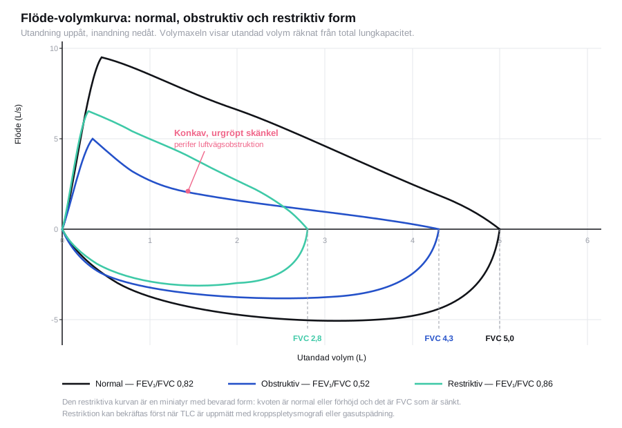
A restrictive pattern on spirometry is only a suspicion — restriction can be confirmed only when the TLC has been measured. A reduced FVC with a normal ratio can equally well be due to incomplete expiration.
Modern guidelines prefer to grade with a z-score rather than with the percentage of predicted: z ≥ −1.65 is normal, −1.65 to −2.5 mild, −2.51 to −4.0 moderate and below −4.0 severe. The z-score takes into account that the spread in the reference population varies with age and height, which the percentage of predicted does not.
The shape of the flow-volume loop
| Shape of the curve | Interpretation |
|---|---|
| A concave, "scooped-out" expiratory limb | Peripheral obstruction — asthma, COPD |
| A miniature curve with preserved shape | Restriction |
| Rectangular, flattened both in inspiration and expiration | Fixed central airway obstruction, for example tracheal stenosis |
| A flattened inspiratory limb | Variable extrathoracic obstruction — vocal cord dysfunction, goitre |
| A flattened expiratory limb | Variable intrathoracic obstruction |
Vocal cord dysfunction is often mistaken for difficult asthma. Always look at the inspiratory part of the loop before treatment is escalated further.
Complications
Spirometry is a safe investigation, but the forced manoeuvre can provoke coughing, dizziness, headache, syncope (from a reduced venous return), bronchospasm, incontinence and — very rarely — pneumothorax or retinal haemorrhage. Have the patient seated, stop if there is dizziness, and have a short-acting bronchodilator available.
Aftercare and follow-up
Print the curves and keep them — the shape carries information that the numbers do not, and comparison over time requires that the old curves are still available. State in the report whether the patient was on medication, how many acceptable manoeuvres were achieved, and whether a reversibility test was performed.
In newly diagnosed COPD: check the spirometry annually. In asthma: after 3 months of treatment and at least annually thereafter. A normal result does not exclude asthma — variable obstruction may require a PEF chart over two weeks, an exercise test or a methacholine challenge.
Common pitfalls
- Too short an expiration gives a falsely low FVC and thereby a falsely normal ratio — the single commonest reason for COPD being missed in primary care.
- A fixed ratio threshold of 0.70 over-diagnoses COPD in older people and under-diagnoses it in the young. Use the lower limit of normal where the spirometer calculates it.
- A diagnosis made without bronchodilatation. The diagnosis of COPD requires persistent obstruction after a bronchodilator.
- A leak at the mouthpiece in patients with dentures — check that the lips seal tightly, and ask the patient to keep the denture in.
- Restriction reported as established without measurement of lung volumes.
- Reversibility interpreted as synonymous with asthma. Many patients with COPD show reversibility, and patients with asthma can have normal spirometry between attacks.
- No calibration. An uncalibrated spirometer can be several per cent out, which is enough to move a patient across a diagnostic threshold.

