
A cast should hold the fracture still and do nothing else. The two ways in which casting harms the patient are that it is too tight — the swelling comes after the injury, not before — and that an edge or a fold presses on a bony prominence until the skin breaks down. An acute, fresh injury is therefore immobilised in a backslab or in a split cast, never in a closed circumferential cast, and the patient must leave with clear information about the symptoms that should make them seek help immediately.
Indications
- An undisplaced or reduced fracture to be treated conservatively.
- Immobilisation after reduction of a dislocation.
- Immobilisation of a ligament injury, a tendon injury, or after suturing over a joint.
- Pain relief and stabilisation for transfer in a suspected fracture.
- Clinical suspicion of a scaphoid fracture despite a normal radiograph.
Contraindications
- An open fracture or a wound that needs to be inspected — use a windowed splint or external fixation.
- Established or impending compartment syndrome.
- An unstable fracture requiring operative fixation.
- A circumferential cast in a fresh injury, before surgery, or while swelling is progressing.
- Take care in impaired sensation (neuropathy, spinal cord injury) — the patient does not feel the pressure sore.
Preparation and equipment
| Material | Comment |
|---|---|
| Stockinette | Innermost against the skin, protects against the edges of the cast |
| Padding | The wadding that protects the skin and the bony prominences |
| Plaster bandages and plaster slabs (calcium sulfate) | Mouldable, cheap, heavier, less strong, takes longer to set |
| Synthetic cast material (fibreglass) | Lighter, stronger, radiolucent, tolerates moisture better, more expensive and stiffer to mould |
| Elastic bandage | Holds the slab in place; wound on without tension |
| Tepid water, plastic gloves, plaster scissors, cast saw, protective sheeting | Hot water gives faster setting and more heat production — a risk of burns |
Document the distal neurovascular status before casting: sensation, motor function, pulses and capillary refill. Check the skin, and remove rings and jewellery distal to the injury before the swelling develops.
Procedure
- Place the joint at the angle the treatment requires before the cast is applied, and hold that position until the cast has set.
- Pull on the stockinette so that it extends a few centimetres beyond the planned ends of the cast.
- Pad evenly, 2–3 layers with about 50 per cent overlap, and add extra padding over bony prominences, at the ends of the cast and where the edge of the slab will lie. Uneven padding, folds and lumps are the commonest cause of pressure sores.
- Dip the plaster in tepid water and squeeze out the excess without wringing.
- Put the slab in place and mould with the whole palm, never with the fingertips — fingerprints in the plaster become pressure points against the skin.
- Fold the ends of the stockinette back over the edge of the cast and smooth the edges.
- Secure with an elastic bandage in even turns without tension.
- Check that the slab ends where it should: the metacarpophalangeal joints must be able to flex freely, the thumb must be free unless it is to be immobilised, and the elbow must be able to flex if it is to be free.
- Check the distal neurovascular status again once the cast has set, and document it.
- Elevate the limb and inform the patient.
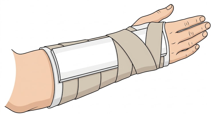
Backslab or circumferential cast
- A dorsal backslab or another half-slab for a fresh injury, acute swelling, after reduction and before surgery. The slab allows the volume to increase.
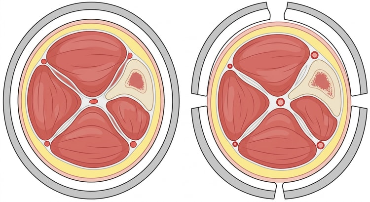
- A circumferential cast gives better stability, but only once the swelling has settled, often after about a week. A circumferential cast applied acutely must be split through its entire thickness, including the padding and the stockinette, right down to the skin — a cast split only through the plaster layer does not relieve the pressure.
Common splints and casts
| Injury | Immobilisation | Comment |
|---|---|---|
| Distal radius fracture, acute | Dorsal backslab, forearm–wrist | Circumferential cast after the swelling phase |
| Suspected scaphoid fracture | Scaphoid cast including the base of the thumb | Reassessment after 10–14 days |
| Finger fracture or finger dislocation | Finger splint or buddy taping | Do not immobilise a finger longer than necessary; risk of stiffness |
| Elbow dislocation after reduction | Dorsal backslab, upper arm–forearm, elbow at 90 degrees | For a short period, then early mobilisation |
| Ankle fracture, acute | Posterior below-knee backslab, ankle at 90 degrees | Circumferential below-knee cast later |
| Marked ankle sprain | Splint or orthosis | An orthosis is usually preferable |
| Metatarsal fracture | Below-knee cast with a heel or a rigid sole | Weight-bearing as directed by the orthopaedic surgeon |
| Achilles tendon rupture | Below-knee cast in equinus, or an orthosis with heel wedges | The angle is changed in stages |
Casting times and weight-bearing regimens vary between regions and units and are governed by the fracture type — follow the local care programme and the orthopaedic surgeon's instructions, not a general rule of thumb.
Compartment syndrome
The feared complication. The pressure in a muscle compartment rises until perfusion ceases, and the muscle necrosis is irreversible within hours.
- Pain that is disproportionately severe and increasing despite adequate analgesia is the first and most important sign.
- Pain on passive stretching of the muscles in the compartment.
- A tense, hard and tender compartment.
- Paraesthesiae and numbness distally.
- A preserved pulse and normal capillary refill do not exclude compartment syndrome — pulselessness is a late and unreliable sign.
Action when it is suspected: split the cast immediately through all layers down to the skin and spread it open, place the limb at the level of the heart — not higher, since elevation lowers the perfusion pressure, and contact the on-call orthopaedic surgeon urgently. Fasciotomy is the definitive treatment.
Complications
- Compartment syndrome.
- Pressure sores over bony prominences. Common sites: the heel, the malleoli, the head of the fibula, the olecranon, the knuckles, the radial styloid and the base of the thumb.
- Pressure on the common peroneal nerve at the head of the fibula causes foot drop — check dorsiflexion of the foot with every below-knee cast.
- Circulatory compromise with cyanosis, swelling and coldness distally.
- Burns from water that is too hot, or from too many layers setting at the same time.
- Joint stiffness and muscle atrophy from prolonged immobilisation.
- Venous thromboembolism when the lower limb is immobilised.
- Itching, eczema and maceration under the cast, often after it has become wet.
Aftercare and follow-up
The patient must be given both verbal and written information. Seek help immediately if:
- The pain increases despite elevation and analgesia.
- There is numbness, tingling or weakness distally.
- The fingers or toes become blue, white, cold or markedly swollen.
- The cast feels too tight, or conversely becomes loose.
- There is a discharging patch, a smell or fever.
- Something has fallen inside the cast.
Other advice: elevation for the first few days, move the fingers and toes regularly, keep the cast dry, never push anything inside to scratch, and never cut or saw the cast yourself.
A follow-up appointment according to the fracture type — a radiograph after about a week in fractures at risk of displacement, clinical review and a change of cast once the swelling has settled, and reassessment after 10–14 days in a suspected scaphoid fracture.
Common pitfalls
- A closed circumferential cast in a fresh injury.
- A cast split only through the plaster layer, while the padding and the stockinette still encircle the limb.
- Uneven or too thin padding, or padding with folds in it.
- Moulding with the fingertips, which creates pressure points.
- A slab that ends too distally and blocks the metacarpophalangeal joints — this leads to stiffness.
- No documented distal neurovascular status before and after casting.
- Elevation above the level of the heart in suspected compartment syndrome, which worsens the perfusion.
- The patient goes home without warning information.

