The electrical axis of the heart (heart axis)
Although often ignored, assessment of the electrical axis is an integral part of ECG interpretation. The electrical axis reflects the average direction of ventricular depolarization during ventricular contraction. The direction of the depolarization (and thus the electrical axis) is generally alongside the hearts longitudinal axis (to the left and downwards). Figure 38 shows the coordinate system where the green area displays the range of normal heart axis.
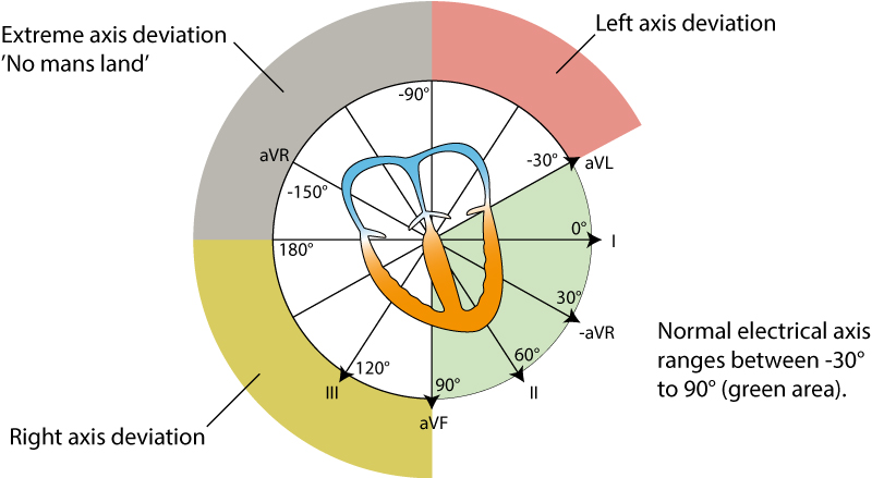
As evident from the figure, the normal heart axis is between –30° and 90°. If the axis is more positive than 90° it is referred to as right axis deviation. If the axis is more negative than –30° it is referred to as left axis deviation. The axis is calculated (to the nearest degree) by the ECG machine. The axis can also be approximated manually by judging the net direction of the QRS complex in leads I and II. The following rules apply:
- Normal axis: Net positive QRS complex in leads I and II.
- Right axis deviation: Net negative QRS complex in lead I but positive in lead II.
- Left axis deviation: Net positive QRS complex in lead I but negative in lead II.
- Extreme axis deviation (–90°to 180°): Net negative QRS complex in leads I and II.
Heart axis deviation: right axis deviation (RAD) and left axis deviation (LAD)
Causes of right axis deviation
Normal in newborns. Right ventricular hypertrophy. Acute cor pulmonale (pulmonary embolism). Chronic cor pulmonale (COPD, pulmonary hypertension, pulmonary valve stenosis). Lateral ventricular infarction. Pre-excitation. Switched arm electrodes (negative P and QRS-T in lead I). Situs inversus. Left posterior fascicular block is diagnosed when the axis is between 90° and 180° with rS complex in I and aVL as well as qR complex in III and aVF (with QRS duration <0.12 seconds), provided that other causes of right axis deviation have been excluded.
Causes of left axis deviation
Left bundle branch block. Left ventricular hypertrophy. Inferior infarction. Pre-excitation. Left anterior fascicular block is diagnosed if the axis is between -45° and 90° with qR complex in aVL and QRS duration is 0,12 s, provided that other causes of left axis deviation have been excluded.
Causes of extreme axis deviation
Rare. Most likely due to misplaced limb electrodes. If the rhythm is tachycardia with wide QRS complexes, then ventricular tachycardia is the most likely cause.
This article is part of the comprehensive chapter: How to read and recognize the normal ECG

