Reference (normal) values for echocardiography

Aorta

Normal values for aorta in 2D echocardiography

 Normal intervalNormal interval, adjusted
Aortic annulus20-31 mm12-14 mm/m2
Sinus valsalva29-45 mm15-20 mm/m2
Sinotubular junction22-36 mm13-17 mm/m2
Ascending aorta22-36 mm13-17 mm/m2
Aortic arch22-36 mm 
Descending aorta20-30 mm 

​Adjustment refers to adjusting for body surface area (BSA).

BSA: m2

Män

Sinus valsalva

Min: mm

Max: mm

Aorta ascendens

Min: mm

Max: mm


Kvinnor

Sinus valsalva

Min: mm

Max: mm

Aorta ascendens

Min: mm

Max: mm


Min och max anger 95% konfidensintervall.
Min= 5:e percentil.
Max = 95:e percentil.

Data baserad på linjär regressionsmodell av Biaggi et al (Gender, age, and body surface area are the major determinants of ascending aorta dimensions in subjects with apparently normal echocardiograms, J Am Soc Echocardiogr. 2009 Jun;22(6):720-5).

Standardized values for aorta (mean ± SD)

 Aortic annulusSinus valsalva
AgeWomenMenWomenMen
<3011.4 ± 1.111.4 ± 1.215.5 ± 1.515.3 ± 1.8
30–3911.7 ± 1.011.4 ± 1.116.5 ± 1.615.7 ± 1.3*
40–4911.7 ± 1.011.5 ± 0.916.8 ± 1.416.6 ± 1.5
50–5912.0 ± 0.911.5 ± 0.917.9 ± 1.616.8 ± 1.7*
60–6911.7 ± 1.112.1 ± 1.017.5 ± 1.717.8 ± 1.6
>7011.6 ± 1.112.0 ± 1.018.2 ± 1.918.1 ± 1.7
All11.7 ± 1.011.6 ± 1.017.0 ± 1.916.6 ± 1.8
 Sinotubular junctionAscending aorta
AgeWomenMenWomenMen
<3013.5 ± 1.412.7 ± 2.315.1 ± 1.614.3 ± 1.6*
30–3914.3 ± 1.513.3 ± 1.316.4 ± 1.814.5 ± 1.4*
40–4914.6 ± 1.214.1 ± 1.5*16.4 ± 1.415.5 ± 1.5*
50–5915.7 ± 1.314.1 ± 1.518.0 ± 1.416.2 ± 1.6*
60–6914.7 ± 1.615.1 ± 1.6*17.6 ± 1.717.3 ± 1.7
>7015.5 ± 2.014.7 ± 1.618.2 ± 3.317.5 ± 1.4
All14.7 ± 1.713.9 ± 1.816.9 ± 2.315.7 ± 1.8
 Aortic arch
AgeWomenMen
<3010.7 ± 1.010.1 ± 1.3*
30–3911.6 ± 1.610.8 ± 1.3*
40–4911.5 ± 1.211.3 ± 1.3
50–5912.7 ± 1.111.3 ± 1.2*
60–6912.5 ± 1.412.0 ± 1.1
>7012.7 ± 1.212.2 ± 1.3
All11.9 ± 1.511.2 ± 1.4

Aortic valve regurgitation, normal values

Primary parameters

 Mild regurgitationModerate regurgitationSevere regurgitation
Vena contracta (cm)< 0,30,3-0,6> 0,6
PHT (pressure half time) – CW doppler> 500200-500< 200
Diastolic flow reversal – PW doppler aorta descendensif present, early diastolicearly diastolicholodiastolic

Quantification of jet

 Mild regurgitationModerate regurgitationSevere regurgitation
Jet-width / LVOT-width< 0,250,25-0,64> 0,64
Jet-area / LVOT-area< 0,050,05-0,59> 0,59

Qualitative parameters

 Mild regurgitationModerate regurgitationSevere regurgitation
Jet areasmallvariablelarge if central, variable if excentric
Coaptation defectmildmoderatesevere
CW-profile aortic regurgitationweakvariabledense
Size left atriumnormalnormal or enlargedenlarged
Size left ventriclenormalnormal or enlargedenlarged

Additional PISA-parameters

 Mild regurgitationModerate regurgitationSevere regurgitation
EROA (cm²)< 0,100,10-0,29> 0,29
Regurgitation volume (ml/beat)< 3030-59> 59
Regurgitation fraction (%)< 3030-49> 49

EROA = (CSA*v)/v(maxMR) {{ Flödeshastighet = CSA*v = 6.28*r2*v(aliasing)

References

1. Zoghbi, William A.; Enriquez-Sarano, Maurice; Foster, Elyse; Grayburn, Paul A.; Kraft, Carol D.; Levine, Robert A. et al. (2003): Recommendations for evaluation of the severity of native valvular regurgitation with two-dimensional and Doppler echocardiography. In Journal of the American Society of Echocardiography : official publication of the American Society of Echocardiography 16 (7), pp. 777–802. DOI: 10.1016/S0894-7317(03)00335-3.

2. Zoghbi, William A.; Adams, David; Bonow, Robert O.; Enriquez-Sarano, Maurice; Foster, Elyse; Grayburn, Paul A. et al. (2017): Recommendations for Noninvasive Evaluation of Native Valvular Regurgitation. A Report from the American Society of Echocardiography Developed in Collaboration with the Society for Cardiovascular Magnetic Resonance. In Journal of the American Society of Echocardiography : official publication of the American Society of Echocardiography 30 (4), pp. 303–371. DOI: 10.1016/j.echo.2017.01.007.

Aortic valve stenosis

Disease defining parameters

 Mild stenosisModerate stenosisSevere stenosis
Valve area (cm²)> 1,51,0-1,5< 1,0
Normalized valve area (cm²/m²)>0,850,6-0,85< 0,6

Measured parameters

 Aortic sclerosisMild stenosisModerate stenosisSevere stenosis
Mean gradient (mmHg) < 2020-40> 40
Peak velocity (m/s)1,8-2,52,6-2,93,0-4,0> 4,0
Peak gradient (mmHg) < 3535-65> 65
Velocity ratio (LVOT / aortic valve) > 0,50,5-0,25< 0,25

References

1. Baumgartner, Helmut; Hung, Judy; Bermejo, Javier; Chambers, John B.; Evangelista, Arturo; Griffin, Brian P. et al. (2009): Echocardiographic assessment of valve stenosis: EAE/ASE recommendations for clinical practice. In Journal of the American Society of Echocardiography : official publication of the American Society of Echocardiography 22 (1), 1-23; quiz 101-2. DOI: 10.1016/j.echo.2008.11.029.

2. Baumgartner, Helmut; Hung, Judy; Bermejo, Javier; Chambers, John B.; Evangelista, Arturo; Griffin, Brian P. et al. (2009): Echocardiographic assessment of valve stenosis: EAE/ASE recommendations for clinical practice. In European journal of echocardiography : the journal of the Working Group on Echocardiography of the European Society of Cardiology 10 (1), pp. 1–25. DOI: 10.1093/ejechocard/jen303.

3. Vahanian, Alec; Alfieri, Ottavio; Andreotti, Felicita; Antunes, Manuel J.; Barón-Esquivias, Gonzalo; Baumgartner, Helmut et al. (2012): Guidelines on the management of valvular heart disease (version 2012). In European heart journal 33 (19), pp. 2451–2496. DOI: 10.1093/eurheartj/ehs109.

4. Baumgartner, Helmut; Hung, Judy; Bermejo, Javier; Chambers, John B.; Edvardsen, Thor; Goldstein, Steven et al. (2017): Recommendations on the Echocardiographic Assessment of Aortic Valve Stenosis. A Focused Update from the European Association of Cardiovascular Imaging and the American Society of Echocardiography. In Journal of the American Society of Echocardiography : official publication of the American Society of Echocardiography 30 (4), pp. 372–392. DOI: 10.1016/j.echo.2017.02.009.

Left and right ventricle

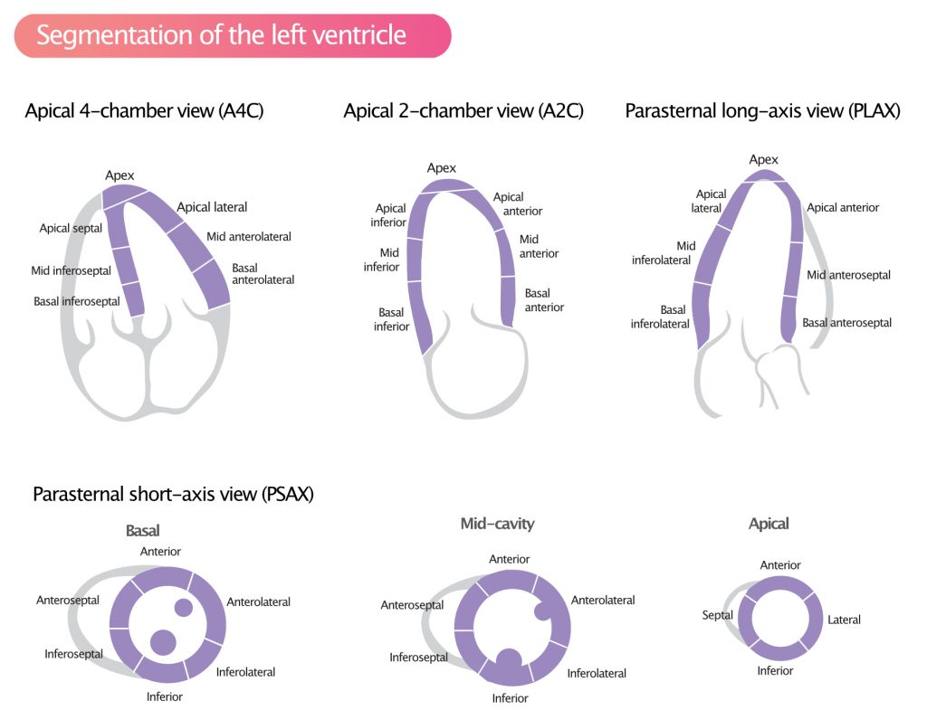
Visual assessment of regional wall motion (left ventricle)

TYPE OF MOTIONDEFINITIONWALL MOTION SCORE
NormalNormal thickening (typically >30% thickening from end-diastole to end-systole).1
HypokinesiaReduced thickening (typically 10–30% thickening from end-diastole to end-systole)2
AkinesiaMarkedly reduced, or no thickening (<10%)3
DyskinesiaParadoxical thinning and/or outward directed motion during systole4
AneurysmaticDiastolic deformation5

Recommended by American Society for Echocardiography (J Am Soc Echocardiogr 18:1440-1463, 2005).

Reference values for left ventricular mass and geometry

 Women   Men   
 ReferenceMildy abnormalModerately abnormalSeverely abnormalReferenceMildly abnormalModerately abnormalSeverely abnormal
LINEAR METHOD        
LV mass, g67–162163–186187–210>21188–224225–258259–292>293
LV mass/BSA, g/m243–9596–108109–121> 12249–115116–131132–148> 149
LV mass/height, g/m41–99100–115116–128>12952–126127–144145–162>163
LV mass/height 2,7 , g/m2,718–4445–5152–58>5920–4849–5556–63>64
Relative wall thickness, cm0.22–0.420.43–0.470.48–0.52>0.530.24–0.420.43–0.460.47–0.51>0.52
Septal thickness, cm0.6–0.91.0–1.21.3–1.5> 1.60.6–1.01.1–1.31.4–1.6> 1.7
Posterior wall thickness, cm0.6–0.91.0–1.21.3–1.5> 1.60.6–1.01.1–1.31.4–1.6> 1.7
2D METOD        
LV mass, g66–150151–171172–182> 19396–200201–227228–254> 255
LV mass/BSA, g/m244–8889–100101–112> 11350–102103–116117–130> 131

BSA, Body surface area; LV, left ventricular; 2D, 2-dimensional.

Referensvärden för vänster kammares storlek

 Women   Men   
 Normal rangeMildly abnormalModerately abnormalSeverely abnormalNormal rangeMildly abnormalModerately abnormalSeverely abnormal
LV DIMENSIONS        
LV diastolic diameter3.9–5.35.4–5.75.8–6.1>6.24.2–5.96.0–6.36.4–6.8>6.9
LV diastolic diameter/BSA, cm/m 22.4–3.23.3–3.43.5–3.7>3.82.2–3.13.2–3.43.5–3.6>3.7
LV diastolic diameter/height, cm/m2.5–3.23.3–3.43.5–3.6>3.72.4–3.33.4–3.53.6–3.7>3.8
LV VOLUME        
LV diastolic volume, mL56–104105–117118–130>13167–155156–178179–201>201
LV diastolic volume/BSA, mL/m 235–7576–8687–96> 9735–7576–8687–96> 97
LV systolic volume, mL19–4950–5960–69>7022–5859–7071–82>83
LV systolic volume/BSA, mL/m 212–3031–3637–42> 4312–3031–3637–42> 43

BSA, body surface area; LV, left ventricular.

Reference values for left ventricular function (ejection fraction)

 Women   Men   
 NormalMildly abnormalModerately abnormalSeverely abnormalNormalMildly abnormalModerately abnormalSeverely abnormal
LINEAR METHOD        
Endocardial fractional shortening, %27–4522–2617–21<1625–4320–2415–19<14
Midwall fractional shortening, %15–2313–1411–12<1014–2212–1310–11<10
2D METOD        
Ejection fraction, %>5545–5430–44<30>5545–5430–44<30

2D, Two-dimensional.

Classification of ejection fraction

 NormalMildly abnormalModerately abnormalSeverely abnormal
Ejection fraction (%), biplan, males52-7241-5130-40<30
Ejection fraction (%), biplan, females54-7441-5330-40<30

References

1. Lang, Roberto M.; Badano, Luigi P.; Mor-Avi, Victor; Afilalo, Jonathan; Armstrong, Anderson; Ernande, Laura et al. (2015): Recommendations for cardiac chamber quantification by echocardiography in adults: an update from the American Society of Echocardiography and the European Association of Cardiovascular Imaging. In European heart journal cardiovascular Imaging 16 (3), pp. 233–270. DOI: 10.1093/ehjci/jev014.

2. Lang, Roberto M.; Bierig, Michelle; Devereux, Richard B.; Flachskampf, Frank A.; Foster, Elyse; Pellikka, Patricia A. et al. (2005): Recommendations for chamber quantification: a report from the American Society of Echocardiography’s Guidelines and Standards Committee and the Chamber Quantification Writing Group, developed in conjunction with the European Association of Echocardiography, a branch of the European Society of Cardiology. In Journal of the American Society of Echocardiography : official publication of the American Society of Echocardiography 18 (12), pp. 1440–1463. DOI: 10.1016/j.echo.2005.10.005. –>Pubmed-Link
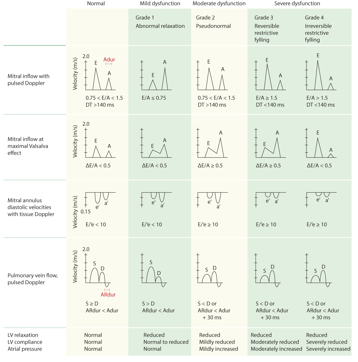
CriteriaNormal, youngNormal, adultAbnormal relaxation (grade 1 dysfunction)Pseudonormal (grade 2 dysfunction)Reversible restrictive filling (grade 3 dysfunction)Irreversible restrictive filling (grade 4 dysfunction)
E/A-ratio1–21–2<11–1.5 (not reversed with Valsalva maneuver)>1.51.5–2.0 (not reversed with Valsalva maneuver)
Deceleration time (DT), ms<240150–240≥240150–200<150<150
IVRT (ms)70–9070–90>90<90<70<70
S/D-ratio<1≥1≥1<1<1<1
ARdur – Adur (ms)≥30≤0≤0 or ≥30≥30≥30≥30
AR speed (cm/s)<35<35<35≥35≥35≥35
e’ speed (cm/s)>10>8<8<8<8<8

 Normal intervalMildly abnormalModerately abnormalSeverely abnormal
RV DIMENSIONS    
Basal RV diameter (RVD 1), cm2.0–2.82.9–3.33.4–3.8>3.9
Mid RV diameter (RVD 2), cm2.7–3.33.4–3.73.8–4.1>4.2
Base-to-apex length (RVD 3), cm7.1–7.98.0–8.58.6–9.1>9.2
RVOT DIAMETER    
Above aortic valve (RVOT 1), cm2.5–2.93.0–3.23.3–3.5>3.6
Above pulmonic valve (RVOT 2), cm1.7–2.32.4–2.72.8–3.1>3.2
PA DIAMETER    
Below pulmonic valve (PA 1), cm1.5–2.12.2–2.52.6–2.9>3.0
RV SIZE AND FUNCTION (MEASURED IN A4C)    
RV diastolic area, cm211–2829–3233–37>38
RV systolic area, cm27.5–1617–1920–22>23
RV fractional area change, %32–6025–3118–24<17

Left and right atrium

Reference values for atrial size and volume

 Women   Men   
 Normal rangeMildly abnormalModerately abnormalSeverely abnormalNormal rangeMildly abnormalModerately abnormalSeverely abnormal
ATRIAL DIMENSIONS        
LA diameter, cm2.7–3.83.9–4.24.3–4.6>4.73.0–4.04.1–4.64.7–5.2>5.2
LA diameter/BSA, cm/m21.5–2.32.4–2.62.7–2.9>3.01.5–2.32.4–2.62.7–2.9>3.0
RA minor-axis dimension, cm2.9–4.54.6–4.95.0–5.4>5.52.9–4.54.6–4.95.0–5.4>5.5
RA minor-axis dimension/BSA, cm/m21.7–2.52.6–2.82.9–3.1>3.21.7–2.52.6–2.82.9–3.1>3.2
ATRIAL AREA        
LA area, cm2<2020–3030–40>40<2020–3030–40>40
ATRIAL VOLUMES        
LA volume, mL22–5253–6263–72>7318–5859–6869–78>79
LA volume/BSA, mL/m222 ± 629–3334–39>4022 ± 629–3334–39>40

​BSA, Body surface area; LA, left atrial; RA, right atrial.

Mitral valve

Mitral valve regurgitation

Primary parameters

 Mild regurgiationModerate regurgitationSevere regurgitation
Vena contracta (cm)< 0,30,3-0,69> 0,69
EROA (cm²)< 0,200,20-0,39> 0,39
Jet size< 4cm² or < 20% of atrial size4-10 cm² or 20-40% of atrial size> 10 cm² or > 40% of atrial size
Jet orientationcentralvariableexcentric
Pulmonary vein flowS-dominantreduced S-wavesystolic flow reversal,

Qualitative parameters

 Mild regurgiationModerate regurgitationSevere regurgitation
Mitral valve apparatusnormal or mildly abnormaloften abnormalabnormal, possibly flail
CW-profile mitral regurgitationweakvariabledense
Shape CW-profile mitral regurgitationparabolicvariableearly peak, triangular
Size left atriumnormalnormal or dilateddilated
Size left ventriclenormalnormal or dilateddilated

Additional PISA-parameters

 Mild regurgiationModerate regurgitationSevere regurgitation
Regurgitation volume (ml/beat)< 3030-59> 59
Regurgitation volume = EROA x TVI   
Regurgitation fraction (%)< 3030-49> 49

Miscellaneous parameters

 Mild regurgiationModerate regurgitationSevere regurgitation
PW-Doppler mitral valveA dominantvariablehigh E values

​References

1. Zoghbi, William A.; Enriquez-Sarano, Maurice; Foster, Elyse; Grayburn, Paul A.; Kraft, Carol D.; Levine, Robert A. et al. (2003): Recommendations for evaluation of the severity of native valvular regurgitation with two-dimensional and Doppler echocardiography. In Journal of the American Society of Echocardiography : official publication of the American Society of Echocardiography 16 (7), pp. 777–802. DOI: 10.1016/S0894-7317(03)00335-3.

2. Zoghbi, William A.; Adams, David; Bonow, Robert O.; Enriquez-Sarano, Maurice; Foster, Elyse; Grayburn, Paul A. et al. (2017): Recommendations for Noninvasive Evaluation of Native Valvular Regurgitation. A Report from the American Society of Echocardiography Developed in Collaboration with the Society for Cardiovascular Magnetic Resonance. In Journal of the American Society of Echocardiography : official publication of the American Society of Echocardiography 30 (4), pp. 303–371. DOI: 10.1016/j.echo.2017.01.007. –>Pubmed-Link​

Mitral valve stenosis

Primary parameters

 Mild stenosisModerate stenosisSevere stenosis
Valve area> 1,5 cm²1,5 – 1,0 cm²< 1,0 cm²
Mean gradient< 5 mmHg5-10 mmHg> 10 mmHg

Additional parameters

 Mild stenosisModerate stenosisSevere stenosis
Pulmonary artery pressure<3030-50>50

References

1. Baumgartner, Helmut; Hung, Judy; Bermejo, Javier; Chambers, John B.; Evangelista, Arturo; Griffin, Brian P. et al. (2009): Echocardiographic assessment of valve stenosis: EAE/ASE recommendations for clinical practice. In European journal of echocardiography : the journal of the Working Group on Echocardiography of the European Society of Cardiology 10 (1), pp. 1–25. DOI: 10.1093/ejechocard/jen303.

2. Baumgartner, Helmut; Hung, Judy; Bermejo, Javier; Chambers, John B.; Evangelista, Arturo; Griffin, Brian P. et al. (2009): Echocardiographic assessment of valve stenosis: EAE/ASE recommendations for clinical practice. In Journal of the American Society of Echocardiography : official publication of the American Society of Echocardiography 22 (1), 1-23; quiz 101-2. DOI: 10.1016/j.echo.2008.11.029.

3. Vahanian, Alec; Alfieri, Ottavio; Andreotti, Felicita; Antunes, Manuel J.; Barón-Esquivias, Gonzalo; Baumgartner, Helmut et al. (2012): Guidelines on the management of valvular heart disease (version 2012). In European heart journal 33 (19), pp. 2451–2496. DOI: 10.1093/eurheartj/ehs109.

Tricuspid valve

Tricuspid valve regurgitation

Primary parameters

 Mild regurgiationModerate regurgitationSevere regurgitation
PISA radius< 0,60,6-0,9> 0,9
Vena contracta (cm)not defined< 0,7> 0,7
Liver vein flowS-dominantReduced S-wavesystolic flow reversal, jet reaching liver veins

Additional quantitative parameters

 Mild regurgiationModerate regurgitationSevere regurgitation
Jet size< 5cm²5-10 cm²> 10 cm²
size vena cava inferior< 2,1 cm and compressiblein between> 2,1 cm, almost not compressible

Additional qualitative parameters

 Mild regurgiationModerate regurgitationSevere regurgitation
Coaptation defectmildmoderatesevere
Size right ventriclenormalnormal or enlargedenlarged
Size right atriumnormalnormal or enlargedenlarged
CW-profile tricuspid regurgitationweakvariabledense
Shape CW-profile tricuspid regurgitationparabolicvariableearly peak, triangular

References

1. Zoghbi, William A.; Enriquez-Sarano, Maurice; Foster, Elyse; Grayburn, Paul A.; Kraft, Carol D.; Levine, Robert A. et al. (2003): Recommendations for evaluation of the severity of native valvular regurgitation with two-dimensional and Doppler echocardiography. In Journal of the American Society of Echocardiography : official publication of the American Society of Echocardiography 16 (7), pp. 777–802. DOI: 10.1016/S0894-7317(03)00335-3.

2. Zoghbi, William A.; Adams, David; Bonow, Robert O.; Enriquez-Sarano, Maurice; Foster, Elyse; Grayburn, Paul A. et al. (2017): Recommendations for Noninvasive Evaluation of Native Valvular Regurgitation. A Report from the American Society of Echocardiography Developed in Collaboration with the Society for Cardiovascular Magnetic Resonance. In Journal of the American Society of Echocardiography : official publication of the American Society of Echocardiography 30 (4), pp. 303–371. DOI: 10.1016/j.echo.2017.01.007. –>Pubmed-Link

Tricuspid valve stenosis

Mean pressure gradient≥ 5 mmHg
Valve area according to the continuity equation (cm²)≤ 1 cm²
ZTime-velocity-integral of the tricuspid inflow (cm)> 60 cm
Pressure half time (T ½,ms)≥ 190 ms
Right atriumSeverly enlarged
Vena cava inferiorenlarged

References

1. Baumgartner, Helmut; Hung, Judy; Bermejo, Javier; Chambers, John B.; Evangelista, Arturo; Griffin, Brian P. et al. (2009): Echocardiographic assessment of valve stenosis: EAE/ASE recommendations for clinical practice. In European journal of echocardiography : the journal of the Working Group on Echocardiography of the European Society of Cardiology 10 (1), pp. 1–25. DOI: 10.1093/ejechocard/jen303.

2. Baumgartner, Helmut; Hung, Judy; Bermejo, Javier; Chambers, John B.; Evangelista, Arturo; Griffin, Brian P. et al. (2009): Echocardiographic assessment of valve stenosis: EAE/ASE recommendations for clinical practice. In Journal of the American Society of Echocardiography : official publication of the American Society of Echocardiography 22 (1), 1-23; quiz 101-2. DOI: 10.1016/j.echo.2008.11.029. –>Pubmed-Link

Pulmonary valve

Pulmonary valve regurgitation

 Mild regurgiationModerate regurgitationSevere regurgitation
Pulmonary valve morphologynormalnormal/abnormalabnormal
Density CW-Dopplerweak, slow decelerationvariabledense, steep deceleration, early termination of diastolic flow
Jet width colour Dopplersmall,, < 10 mm length, narrow originvariablelarge, wide origin
Ratio pulmonic to aortic flownormalvariablesignificantly increased

References

1. Zoghbi, William A.; Enriquez-Sarano, Maurice; Foster, Elyse; Grayburn, Paul A.; Kraft, Carol D.; Levine, Robert A. et al. (2003): Recommendations for evaluation of the severity of native valvular regurgitation with two-dimensional and Doppler echocardiography. In Journal of the American Society of Echocardiography : official publication of the American Society of Echocardiography 16 (7), pp. 777–802. DOI: 10.1016/S0894-7317(03)00335-3.

2. Zoghbi, William A.; Adams, David; Bonow, Robert O.; Enriquez-Sarano, Maurice; Foster, Elyse; Grayburn, Paul A. et al. (2017): Recommendations for Noninvasive Evaluation of Native Valvular Regurgitation. A Report from the American Society of Echocardiography Developed in Collaboration with the Society for Cardiovascular Magnetic Resonance. In Journal of the American Society of Echocardiography : official publication of the American Society of Echocardiography 30 (4), pp. 303–371. DOI: 10.1016/j.echo.2017.01.007.

3. Lancellotti, Patrizio; Tribouilloy, Christophe; Hagendorff, Andreas; Moura, Luis; Popescu, Bogdan A.; Agricola, Eustachio et al. (2010): European Association of Echocardiography recommendations for the assessment of valvular regurgitation. Part 1: aortic and pulmonary regurgitation (native valve disease). In European journal of echocardiography : the journal of the Working Group on Echocardiography of the European Society of Cardiology 11 (3), pp. 223–244. DOI: 10.1093/ejechocard/jeq030.

Pulmonary valve stenosis

 Mild stenosisModerate stenosisSevere stenosis
Peak velocity (m/s)< 343528> 4
Peak gradient (mmHg)< 3636-64> 64

References

1. Baumgartner, Helmut; Hung, Judy; Bermejo, Javier; Chambers, John B.; Evangelista, Arturo; Griffin, Brian P. et al. (2009): Echocardiographic assessment of valve stenosis: EAE/ASE recommendations for clinical practice. In European journal of echocardiography : the journal of the Working Group on Echocardiography of the European Society of Cardiology 10 (1), pp. 1–25. DOI: 10.1093/ejechocard/jen303.

2. Baumgartner, Helmut; Hung, Judy; Bermejo, Javier; Chambers, John B.; Evangelista, Arturo; Griffin, Brian P. et al. (2009): Echocardiographic assessment of valve stenosis: EAE/ASE recommendations for clinical practice. In Journal of the American Society of Echocardiography : official publication of the American Society of Echocardiography 22 (1), 1-23; quiz 101-2. DOI: 10.1016/j.echo.2008.11.029. –>Pubmed-Link

Figure 1. Standardized myocardial segmentation and nomenclature for echocardiography. The left ventricle is divided into 17 segments for 2D echocardiography. One can identify these segments in multiple views.
Figure 2. Display, on a circumferential polar plot, of the 17 myocardial segments and the recommended nomenclature for tomographic imaging of the heart.
Figure 3. Assignment of the 17 myocardial segments to the territories of the left anterior descending (LAD), right coronary artery (RCA), and the left circumflex coronary artery (LCX).

Join our newsletter and get a free ECG Pocket Guide

ECG pocket guide

Start learning now