
A corticosteroid injection into soft tissue gives rapid but time-limited pain relief and rarely alters the natural history of the disease. In lateral epicondylitis the effect is in fact worse than no treatment at all at one year. The injection is therefore a tool to make rehabilitation possible, not an alternative to it. See the separate guide to joint aspiration and injection.
Indications
| Condition | Injection site | Comment |
|---|---|---|
| Subacromial bursitis, rotator cuff syndrome | The subacromial space | The best documented indication |
| Greater trochanteric pain syndrome (gluteal tendinopathy) | The trochanteric bursa | The effect is chiefly short-term |
| Lateral epicondylitis | At the extensor origin | Consider withholding it; the long-term outcome is worse |
| Medial epicondylitis | At the flexor origin | Avoid the ulnar nerve |
| Carpal tunnel syndrome | The carpal tunnel | An acceptable bridge to surgery |
| De Quervain's tenosynovitis | The first dorsal compartment | A good effect |
| Trigger finger | The tendon sheath at the A1 pulley | A good effect, often curative |
| Plantar fasciitis | The origin of the fascia on the calcaneus | A risk of rupture; use sparingly |
| Prepatellar and olecranon bursitis | The bursa | Exclude septic bursitis first |
Injection is considered when 4–6 weeks of adequate conservative treatment — modification of loading, eccentric exercise, an NSAID, an orthosis — has not given a sufficient effect.
Contraindications
Absolute:
- Cellulitis, a skin wound or other infection over the injection site.
- Bacteraemia or suspected septic bursitis or arthritis.
- A joint prosthesis in the adjacent joint.
- Known hypersensitivity to the preparations.
Relative:
- Poorly controlled diabetes — expect a raised blood glucose for 2–5 days.
- Suspected or known tendon rupture.
- No response to previous injections at the same site.
- More than three injections at the same site within a year.
- The Achilles tendon and the patellar tendon — do not inject into or beside them.
Anticoagulant treatment is not a contraindication to these superficial injections.
Preparation and equipment
- Chlorhexidine in alcohol, an iodine solution or isopropyl alcohol.
- A depot glucocorticoid: triamcinolone hexacetonide (Lederspan 20 mg/mL) or methylprednisolone acetate (Depo-Medrol 40 mg/mL). Triamcinolone acetonide (Kenacort-T) has been out of stock for a long period — check local availability. Betamethasone (Betapred 4 mg/mL) is water-soluble and short-acting, not a depot preparation, and gives a shorter duration of effect.
- Lidocaine 10 mg/mL without adrenaline, 2–5 mL.
- Needles: 21–25 G, 40 mm for subacromial and trochanteric injection; 25–27 G, 25 mm for the carpal tunnel, de Quervain's and trigger finger.
- Syringes of 3, 5 and 10 mL, artery forceps, gauze, a dressing, gloves.
- A cold spray or a lidocaine weal.
| Site | Triamcinolone hexacetonide 20 mg/mL | Methylprednisolone acetate 40 mg/mL | Volume of lidocaine |
|---|---|---|---|
| Subacromial | 20 mg (1 mL) | 20–40 mg (0.5–1 mL) | 4–9 mL |
| Trochanteric | 20 mg (1 mL) | 20–40 mg (0.5–1 mL) | 4–6 mL |
| Epicondylar | 10–20 mg | 20 mg | 1–2 mL |
| Carpal tunnel | 10 mg | 20 mg (or dexamethasone 4–6 mg) | 1–1.5 mL |
| De Quervain's | 10 mg | 20 mg | 1 mL |
| Trigger finger | 10 mg | 10–20 mg | 0.5–1 mL |
| Plantar fascia | 10–20 mg | 20–40 mg | 1–2 mL |
The doses follow the summaries of product characteristics: Lederspan specifies 10–20 mg periarticularly in bursitis, tendinitis, tenosynovitis and epicondylitis, and Depo-Medrol 4–30 mg into a bursa and 4–40 mg in tendinitis and epicondylitis. Higher doses than these add nothing documented but increase the risk of skin and fat atrophy. Lederspan states in addition that the dosing interval should not be shorter than four weeks.
Mix the corticosteroid and the lidocaine in the same syringe. The anaesthetic confirms that the injection has gone to the right place — immediate pain relief is a diagnostic finding — and appears to reduce the risk of a post-injection flare. Lidocaine 10 mg/mL without adrenaline is used; larger volumes are needed subacromially and trochanterically to distribute the dose within the bursa, while tendon sheaths accept less than a millilitre.
Decision pathway
The first step is always to exclude infection. Redness, warmth, fever or fluctuance over a bursa rules out a corticosteroid — in that case aspiration with culture is indicated, not injection.
flowchart TD
  A[Localised soft tissue pain] --> B{Redness, warmth, fever or a systemically unwell patient?}
  B -- Yes --> C[Suspected septic bursitis or arthritis]
  C --> D[Aspiration with culture, CRP and blood cultures. Antibiotics. No corticosteroid injection]
  B -- No --> E{Skin infection, a wound, or a joint prosthesis in the adjacent joint?}
  E -- Yes --> F[Withhold the injection]
  E -- No --> G{Achilles tendon, patellar tendon or suspected tendon rupture?}
  G -- Yes --> H[Do not inject. Refer or arrange ultrasound]
  G -- No --> I{Conservative treatment for 4-6 weeks completed?}
  I -- No --> J[Modify loading, eccentric exercise, an orthosis, analgesia]
  I -- Yes --> K{Three injections at the same site in the past year?}
  K -- Yes --> L[Reconsider the diagnosis. Imaging or referral]
  K -- No --> M[Inject the corticosteroid plus lidocaine. Review after 4-6 weeks]
  M --> N{No effect?}
  N -- Yes --> L
  N -- No --> O[Continue rehabilitation. A further injection no sooner than 3 months later]
Procedure
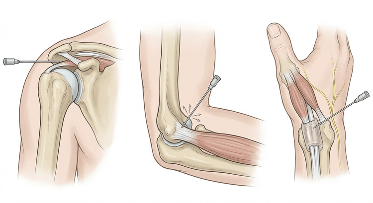
Subacromial injection, lateral approach
- The patient sits with the forearm in the lap so that gravity distracts the humerus and widens the subacromial space. Have the patient grip the edge of the couch and lean towards the opposite side.
- Palpate the lateral edge of the acromion and mark the puncture point just below the edge, above the humeral head.
- Clean the skin, and use a cold spray or a small lidocaine weal.
- Advance the needle horizontally, angled slightly upwards under the acromion, 2–3 cm.
- Aspirate to exclude an intravascular position.
- Inject slowly. The bursa must accept the fluid without resistance. Resistance means tendon or muscle — withdraw the needle slightly and advance it more superiorly.
- Confirm the effect: after a correctly placed injection the pain in the painful arc is almost gone within minutes.
The posterior approach: 2 cm inferior to the posterolateral corner of the acromion, with the needle directed anteriorly towards the coracoid process at an upward angle of 10°.
Trochanteric injection
The patient lies on their side with the painful hip uppermost and the knees flexed. Palpate the point of maximal tenderness over the greater trochanter. Advance the needle perpendicularly until bone contact is reached, withdraw 1–2 mm and inject in a fan-shaped pattern.
Lateral epicondylitis
Palpate the most tender point just distal to the epicondyle, at the origin of extensor carpi radialis brevis. Advance the needle to bone contact, withdraw slightly and inject in several directions in the peritendinous plane. Do not inject into the tendon itself against resistance.
The carpal tunnel
The puncture is made in the wrist crease or 1 cm proximal to it, just ulnar to the palmaris longus tendon. The needle is advanced distally at an angle of 30–45°, 1–1.5 cm. Inject 1.5 mL of a mixture (for example dexamethasone 4 mg/mL 1.5 mL + lidocaine 10 mg/mL 1.5 mL). Stop immediately if there is radiating pain or paraesthesia — the needle is then in the median nerve.
De Quervain's tenosynovitis
The wrist is placed in slight ulnar deviation with the thumb towards the palm. Palpate the first dorsal compartment over the radial styloid process, where abductor pollicis longus and extensor pollicis brevis run in a common tendon sheath. Insert a fine needle at a shallow angle, about 30°, along the axis of the tendons and direct it proximally until the tip lies within the sheath. Inject 1 mL. The fluid should run in without resistance, and a spindle-shaped swelling along the sheath is often seen as it fills.
Two pitfalls: in about a third of people the compartment is divided by a septum, so that only one of the tendons is reached — a failure of effect suggests this and warrants an ultrasound-guided injection. And the superficial branch of the radial nerve lies superficially over the area; radiating pain or numbness on the dorsum of the hand means the needle must be withdrawn.
Trigger finger
The puncture is made over the A1 pulley at the level of the metacarpal head, in the distal palmar crease for the middle and ring fingers. Advance the needle at an angle of 30–45° directed distally until it reaches the tendon; ask the patient to flex the finger gently — if the needle moves with the tendon the tip is within the tendon and must be withdrawn a millimetre or so before the injection is given into the sheath. Inject 0.5–1 mL without resistance.
Complications
| Complication | Comment |
|---|---|
| Post-injection flare | A chemical synovitis within hours, resolving within 48 hours |
| Skin and fat atrophy, depigmentation | From corticosteroid deposited subcutaneously; commoner with superficial injections and in darker skin |
| Tendon rupture | Above all after intratendinous injection and after repeated injections |
| Infection | Rare (< 1/10,000) but serious |
| Hyperglycaemia | For 2–5 days in patients with diabetes — warn them in advance |
| Facial flushing | Common, harmless, 1–2 days |
| Nerve injury | From a misplaced needle, particularly in the carpal tunnel and at the medial epicondyle |
| Vasovagal reaction | Have the patient lie down if in doubt |
Aftercare and follow-up
Relative rest for 1–2 weeks, but no immobilisation — at the shoulder this risks provoking a frozen shoulder. Range-of-movement exercises from day two, progressive loading after two weeks. Ice and paracetamol as required.
Tell the patient to seek help if there is increasing pain after a few hours, fever, or pain that persists beyond 48 hours — these may indicate infection.
Review after 4–6 weeks. If there is no effect at all, the diagnosis should be reconsidered rather than the injection repeated. At most three injections at the same site per year, with an interval of at least three months.
Common pitfalls
- Injecting against resistance — the needle is then in tendon or muscle, and a corticosteroid there weakens the tissue.
- Injection instead of rehabilitation. The effect is short-term; it is the exercise that lasts.
- A corticosteroid in the Achilles or patellar tendon. The risk of rupture makes it indefensible.
- Septic bursitis overlooked — redness, warmth and fever must lead to aspiration and culture, not to a corticosteroid.
- Corticosteroid deposited subcutaneously leaves a visible dimple and a pale patch that the patient remembers for years; empty the needle at depth and withdraw it without injecting on the way out.
- Too high a corticosteroid dose. Periarticularly, 10–20 mg of triamcinolone hexacetonide is enough; more gives no better effect but more atrophy.
- Forgetting to warn a patient with diabetes about the rise in blood glucose.
- Repeated injections without reconsidering the diagnosis.

