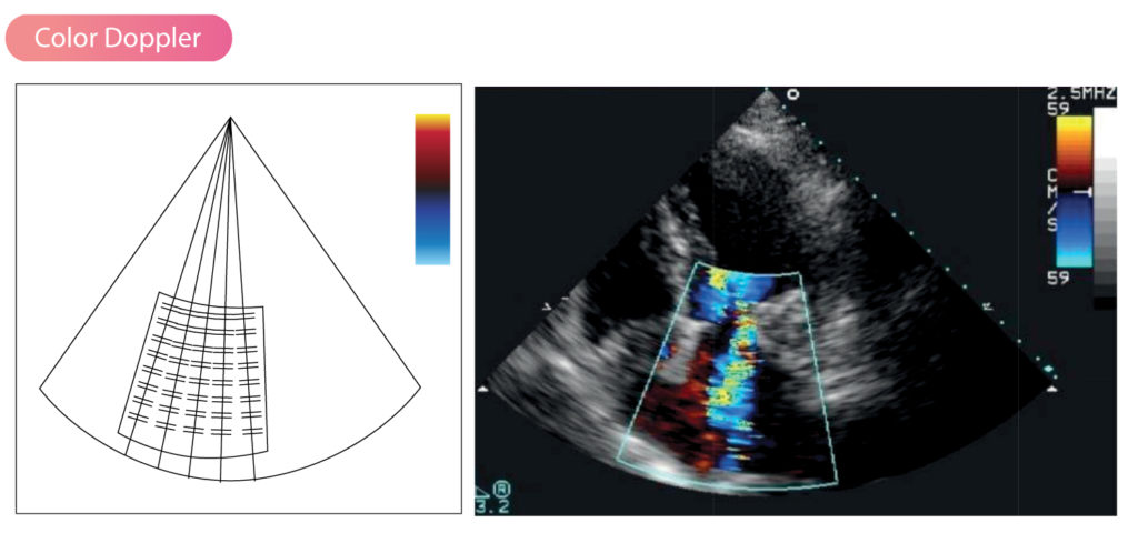
Pulsed Wave Doppler
Pulsed Wave Doppler The pulsed wave Doppler (PW Doppler) sends short pulses of ultrasound and analyzes reflected sound waves between the pulses. This is accomplished…
The Doppler effect When sound waves hit objects some of the sound waves are reflected back to the sound source. If the reflector (i.e the…
M-mode echocardiography M-mode was previously the dominating modality in echocardiography. Although it has now largely been replaced by 2D echocardiography, it is still used in…
Principles of image optimization in echocardiography In order to obtain optimal ultrasound images, it is necessary to adjust several parameters continuously during the examination. Typically,…
Echocardiography in 2D Two-dimensional (2D) ultrasound is the most commonly used modality in echocardiography. The two dimensions presented are width (x axis) and depth (y…
Generating the ultrasound image The ultrasound transducer generates short bursts (pulses) of ultrasound waves. Reflected ultrasound waves are analyzed by the machine during the brief…
The ultrasound transducer & piezoelectric crystals The ultrasound transducer generates ultrasound (ultrasonic) waves. The transducer is held with one hand and its position and angle…
Basic sound and ultrasound physics Unlike light waves, which can propagate through vacuum, sound waves can only propagate through a physical medium. This medium may…
Constrictive pericarditis: the end stage of pericardial inflammation Constrictive pericarditis is the result of chronic inflammation of the pericardium. In principle, all causes of pericarditis…
Hypertrophic cardiomyopathy (HCM): from pathophysiology to echocardiography Hypertrophic cardiomyopathy is a genetic disorder that causes left ventricular hypertrophy under normal loading conditions. Hypertrophic cardiomyopathy should…