
Transcutaneous pacing takes seconds to start and can keep a patient alive until a transvenous lead is in place — but only if it is working, and the commonest source of error is believing that it is. Twitching of the chest muscles and spikes on the screen mean nothing. Capture is verified only when every pacing spike is followed by a wide QRS complex and a pulse that can be felt. The other predictable error is forgetting that pacing hurts a great deal in a conscious patient.
Indications
Immediate (transcutaneous) pacing in bradycardia with haemodynamic compromise — hypotension, shock, impaired consciousness, heart failure, myocardial ischaemia or syncope — that has not responded to atropine:
- Complete heart block (third-degree AV block) and second-degree AV block Mobitz type 2.
- Marked sinus bradycardia, sinus arrest, sick sinus syndrome with long pauses.
- Bradycardia in poisoning with a beta blocker, a calcium channel blocker or digitalis, while awaiting the antidote and specific treatment.
- Bradyasystole during and after cardiac arrest with persisting electrical activity.
- Overdrive pacing in bradycardia-dependent torsades de pointes.
Standby pacing — pads applied but the device not switched on — in second-degree AV block Mobitz type 2, newly discovered complete heart block without current symptoms, alternating bundle branch block in acute anterior infarction, and before drugs or procedures with a known risk of bradycardia.
Transvenous pacing is chosen when the need is more than brief: continuing pacing dependence, inadequate or unachievable transcutaneous capture, or when the patient cannot tolerate the pain of transcutaneous pacing. It is a bridge to a permanent pacemaker or to resolution of the underlying cause.
Contraindications
- Hypothermia with a body temperature below about 30 °C — the hypothermic myocardium is irritable and pacing can precipitate ventricular fibrillation. Prioritise rewarming.
- Bradycardia in a patient with a normal circulation and no symptoms requires no pacing, only monitoring and preparedness.
- Asystole with no electrical activity at all — pacing has no documented benefit there.
- Transcutaneous pacing: burns or wounds in the area where the pads would go.
- Transvenous pacing: the relative contraindications are the same as for a central venous catheter — coagulopathy, infection at the puncture site, known thrombosis in the target vein. With a mechanical tricuspid prosthesis the lead must not be passed through the valve.
The order of management in acute bradycardia
flowchart TD
  A[Bradycardia] --> B{Signs of haemodynamic compromise?<br/>Hypotension, shock, heart failure, ischaemia, impaired consciousness}
  B -- No --> C[Monitor, look for the cause,<br/>apply pacing pads on standby]
  B -- Yes --> D[Oxygen if hypoxic, venous access, ECG monitoring<br/>Stop drugs that cause bradycardia]
  D --> E[Atropine 0.5–1 mg IV, repeated every 3–5 minutes<br/>to a maximum of 3 mg]
  E --> F{Any effect?}
  F -- Yes --> G[Continued monitoring,<br/>investigate the cause and the need for a pacemaker]
  F -- No --> H[Transcutaneous pacing together with<br/>an infusion of isoprenaline, adrenaline or dopamine]
  H --> I{Capture achieved and tolerable?}
  I -- Yes --> J[Continued pacing with analgesia and sedation]
  I -- No --> K[Transvenous pacemaker]
  J --> K
  K --> L[A permanent pacemaker, or the cause corrected]
Atropine is given in doses of 0.5–1 mg intravenously, repeated every three to five minutes to a total maximum of 3 mg. Doses below 0.5 mg can paradoxically worsen the bradycardia and must be avoided. Atropine acts on the sinus node and the AV node and is therefore ineffective — sometimes harmful — in infranodal block with wide QRS complexes; do not delay pacing in order to try yet another dose. Chronotropic support is given as an infusion of isoprenaline, adrenaline or dopamine according to local practice, and does not replace pacing when the patient is markedly unstable.
Transcutaneous pacing
Equipment
- A defibrillator with a pacing function and self-adhesive multifunction pads.
- A separate three-lead ECG cable connected to the patient — the device senses the rhythm through the cable, not through the pads.
- A pulse oximeter, a blood pressure cuff, and an arterial line for prolonged pacing.
- Venous access, oxygen, suction, airway equipment.
- Analgesia and sedation: an opioid (morphine or fentanyl) combined with a benzodiazepine (midazolam or diazepam) is the commonest combination; propofol is used in units with airway expertise.
Procedure
- Dry the skin and shave where necessary. Do not clip the hair — small cuts cause burning pain.
- Position the pads, preferably anteroposteriorly: one anteriorly over the praecordium to the left of the sternum, the other on the back between the left scapula and the spine. The anterolateral position also works and is retained if time is short or the pads are already in place for defibrillation.
- Connect the ECG cable and choose a limb lead with a clear R wave, so that the device can sense the patient's own beats.
- Set demand mode (synchronised) in a patient with an intrinsic rhythm, so that the stimulus does not fall in the vulnerable phase. Fixed-rate mode is used only when there is no intrinsic rhythm at all or when sensing does not work.
- Set the rate. In an unconscious patient, 70–100 beats/minute. In a conscious patient, a lower rate, down towards 40–50, can provide an adequate circulation with less pain while the analgesia takes effect.
- Give analgesia and sedation before the current is increased, if the patient's condition allows.
- Increase the current from zero in steps until capture is achieved. The threshold is often around 70–80 mA but in practice is considerably higher; in an unconscious patient in an emergency one can start straight at 150–200 mA and then reduce.
- Verify capture in two ways. Electrically: every spike must be followed by a wide QRS complex with a discordant T wave. Mechanically: a pulse in the femoral or carotid artery synchronous with the pacing rate, or alternatively a plethysmographic waveform that follows the pacing. Do not palpate the radial artery — the twitching of the arm muscles can feel like a pulse.
- Set the output about 10 % above the threshold and leave it there.
- Check the blood pressure and the level of consciousness, and document the threshold, the rate and the drugs given.
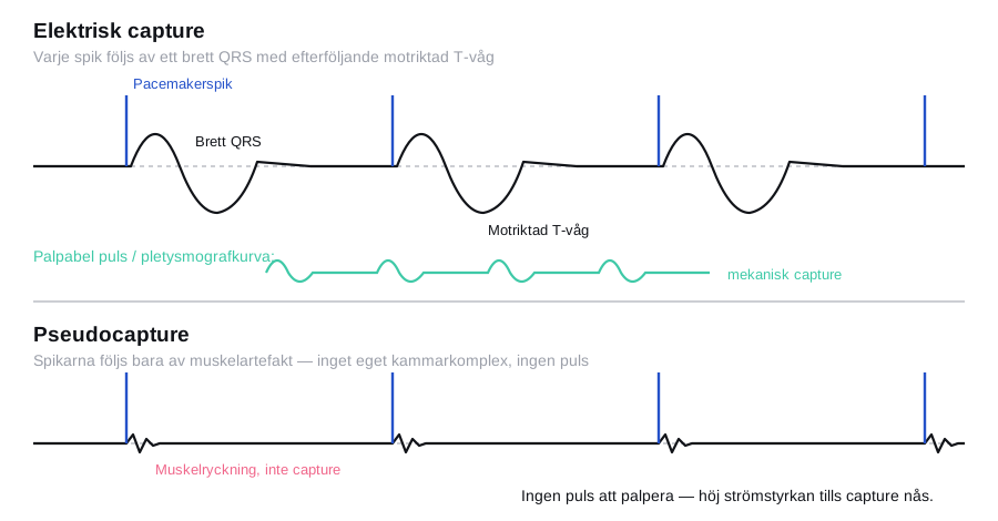
Pain is the rule, not the exception. The conscious patient experiences powerful skeletal muscle contractions and burning discomfort with every stimulus. A patient who pushes your hands away or pulls at the pads is under-treated, not agitated. Transcutaneous pacing is not intended to continue for hours.
Transvenous pacing
Preparation
- Sterile equipment as for a central venous catheter: sterile gown and gloves, chlorhexidine in alcohol, ultrasound with a sterile cover.
- An introducer sheath of a size suited to the lead, and a temporary pacing lead, usually with a balloon for flow-directed advancement.
- An external pulse generator with the battery level checked, and a connecting cable.
- Fluoroscopy where available; in an emergency the lead is instead advanced under ultrasound or ECG guidance.
- Transcutaneous pacing left in place as a safety net throughout the procedure.
Procedure
- Access. The right internal jugular vein gives the straightest route to the right ventricle and is the first choice; the left subclavian vein is an alternative but should be saved for a possible permanent pacemaker. The femoral vein is used when the neck is inaccessible, but it requires fluoroscopy and immobilises the patient. Puncture under ultrasound guidance and insert the sheath with the Seldinger technique.
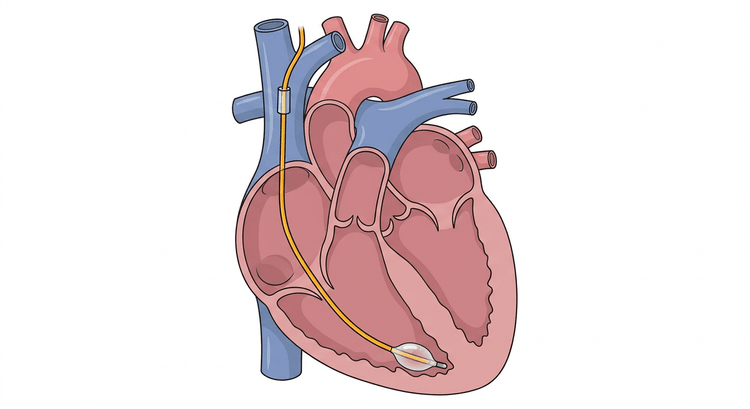
- Advancement. Introduce the lead through the sheath. Inflate the balloon only once the tip has passed the end of the sheath, and let the blood flow carry the lead through the superior vena cava, the right atrium and the tricuspid valve down into the right ventricle.
- Guidance without fluoroscopy: connect the distal pole of the lead to a chest lead on the ECG machine and follow the intracardiac signal pattern — a large P wave in the atrium, a dominant ventricular complex once the valve has been passed, and ST elevation as a sign of contact with the ventricular wall. Alternatively the lead is advanced with the device in pacing mode at a high output; the position is correct when capture occurs with a left bundle branch block-like, superiorly directed ventricular axis.
- The target position is the apex of the right ventricle, with the tip against the trabeculated ventricular wall. Deflate the balloon once the target position has been reached.
- Threshold measurement. Set the rate at least 10 beats above the patient's own and then reduce the current gradually until capture is lost. The threshold must be low, with a guide value below 1 mA (corresponding to about 1 V) — a high threshold means poor contact with the wall and the lead must be repositioned.
- Set the output with a margin: at least two to three times the measured threshold. Thresholds rise over the first few days through local tissue reaction, and an output without a margin means loss of capture during the night.
- Check the sensing. Lower the pacing rate below the patient's own rhythm and check that the device senses the patient's own beats and is inhibited — the sense indicator should flash for every intrinsic QRS. Adjust the sensitivity (mV) until the intrinsic beats are sensed without T waves or interference being counted. A lower mV setting means a higher sensitivity. Undersensing causes stimulation in the vulnerable phase; oversensing causes failure to pace in a pacing-dependent patient.
- Set the final rate, usually 60–80 beats/minute in demand mode.
- Secure the lead with a suture, leave a relief loop and cover with a sterile dressing. Displacement is the commonest complication and is almost always due to inadequate fixation.
- A chest radiograph afterwards to confirm the lead position and exclude a pneumothorax.
- Document the route of access, the depth of insertion, the threshold, the output, the sensitivity and the rate in the notes.
Complications
Transcutaneous pacing:
| Complication | Comment |
|---|---|
| Pain and powerful muscle contractions | The rule in a conscious patient; it requires analgesia and sedation |
| Absent or intermittent capture | Commonest in obesity, emphysema, pericardial effusion and poor skin contact |
| Pseudo-capture mistaken for capture | The most dangerous error — the patient is not being paced at all |
| Skin erythema and superficial burns | With prolonged pacing and a high current |
| Induced ventricular arrhythmia | Rare; the risk increases with fixed-rate mode without demand and in hypothermia |
| A missed diagnosis of ventricular fibrillation | Pacing artefact can conceal underlying VF — always check the pulse |
Transvenous pacing:
| Complication | Comment |
|---|---|
| Lead displacement with loss of capture | The commonest; check the threshold daily and at every alarm |
| Myocardial perforation and tamponade | Feared, uncommon; suspect it with a suddenly rising threshold, chest pain, hiccups, diaphragmatic stimulation or hypotension. Echocardiography immediately |
| Ventricular ectopics and ventricular tachycardia | Common during advancement; they usually stop when the lead is withdrawn slightly |
| Puncture-related: pneumothorax, haemothorax, arterial puncture | Largely avoided with ultrasound-guided puncture |
| Infection, from the puncture site to endocarditis | The risk rises with the dwell time; minimise the days with a lead in place |
| Deep vein thrombosis in the access vein | Particularly with femoral access |
| Tricuspid valve injury | With repeated or forceful manipulation |
| Sensing failure — under- or oversensing | These cause stimulation in the vulnerable phase and failure to pace respectively |
Aftercare and follow-up
- Continuous ECG monitoring for as long as the patient is pacing-dependent, with alarm limits that will detect loss of capture.
- Check the threshold, the sensing and the battery level at least once a day, and at every episode of pacing failure. A rising threshold suggests displacement or tissue reaction.
- Inspect and redress the puncture site according to the local central line procedure; document the number of days in place.
- Keep the settings of the external pulse generator locked or taped over so that they are not changed by accident.
- Consider a permanent pacemaker early. Reversible causes — drugs, hyperkalaemia, ischaemia, hypothyroidism, Lyme disease, infection — must be excluded before a patient receives a permanent device. A temporary lead must not stay in place longer than necessary.
- In inferior myocardial infarction with AV block, the block often resolves within days; in anterior infarction with block the prognosis is worse and a permanent pacemaker is more often necessary.
- Explain to the patient the cause, the driving licence rules after syncope, and what happens next.
Common pitfalls
- Judging capture on the monitor alone. Palpate the femoral or carotid artery — not the radial, where the muscle twitching deceives.
- Confusing muscle twitching with capture. That the chest twitches means only that skeletal muscle is being stimulated.
- Not connecting the ECG cable to the device and believing that the pads are sufficient for sensing.
- Pacing a conscious patient without analgesia and sedation.
- Giving atropine in doses below 0.5 mg, or continuing to give atropine in infranodal block with wide QRS complexes instead of moving on to pacing.
- Leaving transcutaneous pacing running for hours instead of arranging a transvenous lead.
- Accepting a high pacing threshold in transvenous pacing instead of repositioning the lead.
- Setting the output without a margin — the thresholds rise over the first few days.
- Forgetting the sensing check, so that the pacemaker stimulates into the vulnerable phase or is inhibited by interference.
- Forgetting the fixation, the relief loop and the chest radiograph after transvenous insertion.
- Not looking for a reversible cause before a permanent pacemaker is planned.

