
The most serious transfusion accidents rarely stem from laboratory errors but from the wrong blood being given to the wrong patient — a mislabelled tube, or an identity check performed at the nurses' station instead of at the bedside. The check against the patient's own identity immediately before transfusion is the single most important safety measure in the entire chain. In massive haemorrhage a second problem is added: transfusion saves no one whose bleeding is not stopped, and it is the time to haemorrhage control that determines the outcome.
Indications
- Symptomatic anaemia, or a haemoglobin below the applicable transfusion threshold (see below).
- Ongoing bleeding with haemodynamic compromise, whatever the current haemoglobin — haemoglobin falls late in acute haemorrhage and a normal value does not exclude significant blood loss.
- Massive haemorrhage meeting the criteria for activation of the massive transfusion protocol.
- Plasma and platelets: in ongoing bleeding with coagulopathy, or as part of balanced transfusion in massive haemorrhage.
Transfusion thresholds in the stable, non-bleeding patient
| Patient category | Transfusion threshold (Hb) |
|---|---|
| Most stable adult inpatients, including intensive care | 70 g/L |
| Known cardiovascular disease, postoperatively after cardiac surgery | 80 g/L |
| Acute coronary syndrome | 80 g/L; a higher threshold may be considered individually in ongoing ischaemia |
| Upper gastrointestinal bleeding that has been stabilised | 70 g/L — a more liberal strategy gives worse outcomes |
A restrictive strategy is at least equivalent to a liberal one in virtually every group studied. Transfuse one unit at a time and assess the effect clinically and with a repeat haemoglobin before ordering the next unit. In chronic anaemia with a treatable cause — iron deficiency, B12 deficiency, folate deficiency — transfusion is rarely the right measure.
Contraindications
- Anaemia that can be corrected with iron, B12 or folate in a stable patient.
- Transfusion given solely to reach a laboratory value without a clinical question.
- A patient who declines blood transfusion. The decision must be documented and alternatives (iron, tranexamic acid, cell salvage, haemorrhage control) planned in advance.
- A history of a severe transfusion reaction: contact transfusion medicine before transfusing again.
Preparation and equipment
Compatibility testing
| Test | What it involves | When it is used |
|---|---|---|
| Blood grouping | ABO and RhD typing. Requires two samples drawn on two separate occasions with a separate identity check each time | Always, once per patient if no valid result exists |
| Type and screen (BAS test) | Blood group confirmation and antibody screening. If the screen is negative, a unit is selected electronically against the blood group | Standard for planned transfusion in a patient without irregular red cell antibodies |
| Serological crossmatch (MG test) | Serological crossmatching between the recipient's plasma and the red cells of the individual donor unit | When irregular antibodies have been demonstrated, when the screening result is unclear, and otherwise according to local protocol |
Both the type and screen and the serological crossmatch have a limited period of validity — usually about five days — because new antibodies can form after transfusion or pregnancy. Check the validity before ordering blood; an expired type and screen discovered when the patient is already bleeding costs precious time. Exact validity periods and the rules for electronic issue differ between regions — follow the local transfusion medicine regulations.
In life-threatening haemorrhage without a valid blood group, give group O RhD-negative blood (always RhD-negative to women of childbearing age) while awaiting the result.
Equipment
- A large-bore peripheral cannula — at least 1.3 mm (green) for rapid transfusion, preferably two lines in a bleeding patient.
- A transfusion set with a filter. Blood components must not be given through a set without a filter.
- Sodium chloride 9 mg/mL for flushing. No drugs and no other fluid may be given through the same line as the blood component — Ringer's acetate contains calcium, which can cause clot formation in the giving set.
- A blood warmer for rapid transfusion, massive haemorrhage or cold antibodies.
- Monitoring equipment: blood pressure, heart rate, oxygen saturation, temperature.
Procedure
Identity check and transfusion
The check is made at the bedside, by the person starting the transfusion, with the patient present — never in advance at the nurses' station and never by anyone else.
flowchart TD
  A[Blood unit collected to the ward] --> B[Take the unit to the patient's bedside]
  B --> C{The patient states their own name and personal identity number}
  C -- Unable to answer --> D[Check against the identity wristband]
  C -- Answers --> E[Compare with the delivery note and the unit label]
  D --> E
  E --> F{Do the name, identity number, blood group and unit number all match?}
  F -- No --> G[Do not transfuse. Contact the blood bank]
  F -- Yes --> H{Is the unit intact, in date and normal in appearance?}
  H -- No --> G
  H -- Yes --> I[Check vital signs and temperature before starting]
  I --> J[Start slowly and stay with the patient for the first 15 minutes]
  J --> K[Document unit number, time and signature]
Figure 1. The identity check before transfusion. All four items — name, personal identity number, blood group and the unit's identification number — must match both the delivery note and the bag. At the slightest discrepancy the unit is not given.
Transfusion rate:
- Start slowly, about 2 mL/min for the first 15 minutes, and stay with the patient — most life-threatening reactions begin during this phase.
- Thereafter a unit of red cells is normally given over 1–2 hours in a stable patient.
- A unit must be fully transfused within 4 hours of leaving the blood bank's cold chain.
- In heart failure or a high risk of fluid overload: a slower rate, and consider a diuretic between units.
- In ongoing bleeding, the bleeding dictates the rate — the blood is then given as fast as the line allows, through a blood warmer.
Massive haemorrhage
Activate the massive transfusion protocol early in ongoing uncontrolled bleeding with circulatory compromise, when several units are expected to be needed within a short time, or when the estimated bleeding risk is high according to local decision support. Activation must be based on clinical assessment, not on waiting for a haemoglobin value.
flowchart TD
  A[Uncontrolled bleeding with circulatory compromise] --> B[Activate the massive transfusion protocol]
  B --> C[Alert surgery, anaesthesia, interventional radiology and the blood bank simultaneously]
  B --> D[Tranexamic acid 1 g IV, then 1 g over 8 hours]
  B --> E[Two large-bore lines, blood tests including blood gas and coagulation]
  C --> F[Haemorrhage control: surgery, endovascular intervention or compression]
  E --> G[Balanced transfusion: red cells, plasma and platelets 1:1:1]
  G --> H[Blood warmer on everything, active warming of the patient]
  G --> I[Check ionised calcium, give calcium if low]
  H --> J{Bleeding stopped?}
  I --> J
  F --> J
  J -- No --> G
  J -- Yes --> K[Deactivate the protocol, return unused units]
  K --> L[Move to goal-directed replacement guided by laboratory and viscoelastic testing]
Figure 2. The massive transfusion protocol. Activation starts three parallel tracks: haemorrhage control, balanced transfusion, and treatment of hypothermia, acidosis and hypocalcaemia. The protocol is actively deactivated once the bleeding is under control so that unused components can be returned.
Key measures in massive haemorrhage:
- Balanced transfusion in a 1:1:1 ratio of red cells, plasma and platelets, until the bleeding is controlled and goal-directed replacement can take over.
- Tranexamic acid 1 g intravenously as early as possible, followed by 1 g over 8 hours. In trauma the first dose must be given within 3 hours — later administration has not been shown to be of benefit.
- Calcium. Citrate in the blood components binds ionised calcium, and hypocalcaemia impairs both coagulation and myocardial function. Check ionised calcium early and repeatedly, and replace according to local protocol with calcium gluconate or calcium chloride.
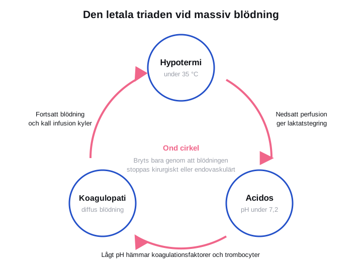
- Avoid large volumes of crystalloid. Fluid dilutes the coagulation factors, cools the patient and worsens the acidosis.
- Permissive hypotension until haemorrhage control may be considered in trauma without head injury — follow the local trauma protocol.
Complications
| Reaction | Typical picture | Management |
|---|---|---|
| Acute haemolytic reaction (ABO incompatibility) | Within minutes: fever, rigors, back and flank pain, hypotension, dark urine, diffuse bleeding. In an anaesthetised patient often only hypotension and diffuse bleeding in the operative field | Stop immediately. Set the giving set and tubing aside, flush with saline through a new set. Fluid and vasopressor, support of renal function, contact transfusion medicine |
| TRALI | Acute hypoxaemia and pulmonary oedema within 6 hours, normal or low filling pressures, often fever | Stop. Oxygen, often ventilatory support. Diuretics have no place — this is not a volume reaction |
| TACO | Dyspnoea, hypertension, raised jugular venous pressure and pulmonary oedema in a patient with cardiac or renal disease, often after several units | Stop or reduce the rate. Sit the patient up, oxygen, diuretics |
| Febrile non-haemolytic reaction | A temperature rise of at least 1 °C and rigors without signs of haemolysis, usually 30–120 minutes into the transfusion | Stop and exclude haemolysis and bacterial contamination. Antipyretics. Transfusion can usually be resumed or continued after assessment |
| Allergic reaction and anaphylaxis | Urticaria and itching; in anaphylaxis laryngeal oedema, bronchospasm and shock | Mild urticaria: antihistamine, slower rate. Anaphylaxis: stop, intramuscular adrenaline (epinephrine), standard anaphylaxis treatment |
| Bacterial contamination | High fever, rigors and shock shortly after starting, usually with a platelet unit | Stop. Blood cultures from the patient and the unit, broad-spectrum antibiotics |
flowchart TD
  A[Symptoms during or shortly after transfusion] --> B[Stop the transfusion immediately]
  B --> C[Keep the line open with sodium chloride through a new giving set]
  C --> D[Re-check the identity against the bag and the delivery note]
  D --> E{Which picture predominates?}
  E -- Fever, rigors, flank pain, dark urine --> F[Suspected haemolysis: samples, fluid, contact transfusion medicine]
  E -- Hypoxia without volume overload --> G[Suspected TRALI: oxygen and ventilatory support, no diuretics]
  E -- Hypoxia with hypertension and raised jugular venous pressure --> H[Suspected TACO: sit the patient up, oxygen, diuretics]
  E -- Urticaria, laryngeal oedema, bronchospasm --> I[Allergic reaction: antihistamine, adrenaline IM in anaphylaxis]
  E -- Fever alone with nothing else --> J[Febrile non-haemolytic reaction once haemolysis has been excluded]
  F --> K[Send the bag and giving set to the blood bank, report the reaction]
  G --> K
  H --> K
  I --> K
  J --> K
Figure 4. Management of a suspected transfusion reaction. The first step is always the same whatever the cause: stop the transfusion and re-check the identity. The bag is never discarded — it must be returned together with the giving set for investigation.
Delayed complications: a delayed haemolytic reaction after 3–14 days with a falling haemoglobin and rising bilirubin, alloimmunisation that complicates future compatibility testing, and iron overload after repeated transfusions.
Aftercare and follow-up
- Check vital signs before starting, after 15 minutes, at the end of the transfusion, and in between according to local protocol.
- Document the unit number, component type, time, volume given, and the prescribing and administering staff in the record and in the transfusion register. Traceability is a statutory requirement.
- Check the haemoglobin no earlier than 15 minutes after the transfusion has finished in a stable patient; in ongoing bleeding the clinical picture governs.
- If a reaction occurs: report it according to local protocol and to transfusion medicine. Give the patient written information about the reaction.
- After massive haemorrhage: continued monitoring for hypocalcaemia, hyperkalaemia, hypothermia, renal failure and respiratory failure, with follow-up of coagulation status.
- Always reconsider the cause of the anaemia. Transfusion treats the value, not the disease.
Common pitfalls
- An identity check made without the patient present. The check must take place at the bedside, against the patient's own statement or their identity wristband.
- Leaving the room during the first 15 minutes. That is when a haemolytic reaction declares itself.
- Ringer's acetate or drugs in the same line as the blood component.
- Waiting to activate the massive transfusion protocol until the haemoglobin result arrives. Haemoglobin falls late — activate on the clinical picture.
- Red cells alone in massive haemorrhage. Without plasma and platelets a dilutional coagulopathy develops.
- Cold transfusion. Unwarmed blood in large volumes is a direct cause of hypothermia and thereby of coagulopathy.
- Forgetting ionised calcium. Citrate-induced hypocalcaemia is common, easy to miss and easy to correct.
- Discarding the bag when a reaction is suspected. Without the bag and giving set the investigation cannot be carried out.
- Giving two units routinely to a stable patient instead of one unit and a fresh assessment.


