
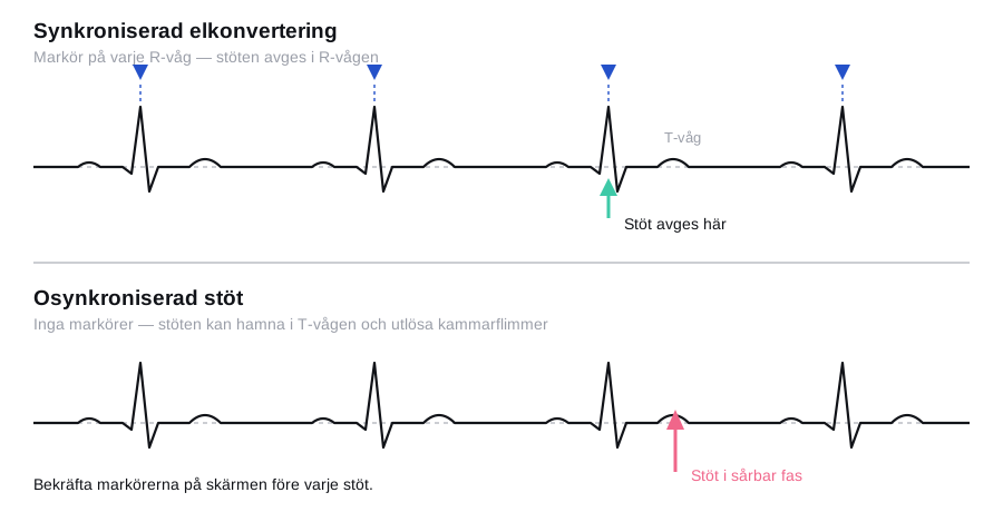
Two things determine whether the procedure is safe: that synchronisation really is switched on when the shock is delivered, and that the thromboembolic risk has been dealt with before the atria begin to contract again. An unsynchronised shock that happens to fall in the T wave can precipitate ventricular fibrillation, and cardioversion of fibrillation of unknown duration without anticoagulation can cause a stroke hours to weeks later. Everything else in the procedure is routine.
Synchronised cardioversion or unsynchronised defibrillation
| Synchronised cardioversion | Unsynchronised defibrillation | |
|---|---|---|
| Rhythm | An organised rhythm with discernible QRS complexes: atrial fibrillation, atrial flutter, SVT, monomorphic VT with a pulse | Ventricular fibrillation, pulseless VT, polymorphic VT (torsades) |
| Timing | The shock is delivered on the R wave | The shock is delivered immediately |
| Why | Avoids the vulnerable phase of the T wave | There is no R wave to synchronise to; sync mode would block the shock entirely |
| The patient | Awake or sedated — requires sedation | Unconscious |
In polymorphic VT, sync must be switched off. The defibrillator finds no stable R wave, and in sync mode no shock at all is then delivered while the patient deteriorates.
Indications
Emergency, irrespective of duration and anticoagulation status:
- Tachyarrhythmia with haemodynamic compromise: hypotension, shock, pulmonary oedema, myocardial ischaemia or a falling level of consciousness.
- Unstable monomorphic VT with a pulse.
- Pre-excitation with atrial fibrillation and a very rapid ventricular rate.
Elective or subacute:
- Symptomatic persistent atrial fibrillation where rhythm control is the aim.
- Atrial flutter, which often converts at low energy and is difficult to terminate pharmacologically.
- Ectopic atrial tachycardia or SVT that has not been terminated by vagal manoeuvres, adenosine or rate-controlling drugs.
- New-onset fibrillation of certain duration below the threshold at which anticoagulation is required.
Contraindications
- Atrial fibrillation or flutter of unknown or excessive duration without adequate anticoagulation or exclusion of thrombus — the most important relative contraindication to elective cardioversion. It does not apply in haemodynamic instability.
- Digitalis toxicity — a risk of refractory ventricular arrhythmias. Therapeutic digoxin levels, by contrast, are no obstacle.
- Untreated hypokalaemia or marked electrolyte disturbance; correct these first before elective cardioversion.
- AVNRT and AVRT that respond to adenosine — try adenosine first in the stable patient.
- Sinus tachycardia and multifocal atrial tachycardia — treat the cause; cardioversion is ineffective.
- Known left atrial thrombus.
- A markedly enlarged left atrium, marked hyperthyroidism or an untreated underlying cause make recurrence all but certain and argue against elective cardioversion.
Anticoagulation before cardioversion
After cardioversion the atrium does not regain its mechanical function until days to weeks have passed — atrial stunning — which means that the thromboembolic risk is greatest after, not during, the cardioversion itself.
- Duration reliably under 48 hours: cardioversion has traditionally been possible without prior anticoagulation. The 2024 ESC guidelines and several Swedish clinical knowledge-support documents have tightened the threshold to 24 hours for patients with risk factors; cardioversion between 24 and 48 hours without ongoing anticoagulation is then reserved for patients with a CHA₂DS₂-VA score of 0–1. Check what applies in your own region.
- Duration above the threshold or unknown: at least three weeks of adequate anticoagulation before cardioversion — an INR ≥ 2.0 checked weekly on warfarin, or documented well-managed DOAC therapy with no missed doses. A single missed DOAC dose makes the pretreatment inadequate and the cardioversion must be postponed.
- Alternative when more rapid cardioversion is needed: transoesophageal echocardiography (TOE) excluding thrombus in the left atrium and the left atrial appendage. Cardiac CT with a late-phase acquisition is used in some units. Anticoagulation must then be started before the cardioversion and continued afterwards.
- After cardioversion: anticoagulation for at least four weeks in every patient, irrespective of duration and method. Thereafter, continued treatment is decided by the CHA₂DS₂-VA score, not by whether sinus rhythm was maintained.
flowchart TD
  A[Atrial fibrillation to be cardioverted] --> B{Haemodynamically unstable?}
  B -- Yes --> C[Immediate synchronised cardioversion<br/>irrespective of duration and anticoagulation]
  C --> H
  B -- No --> D{Ongoing adequate anticoagulation<br/>for at least 3 weeks?}
  D -- Yes --> G[Elective cardioversion can be planned]
  D -- No --> E{Duration reliably below the<br/>local threshold of 24 or 48 hours?}
  E -- Yes --> G
  E -- No or unknown --> F{Is cardioversion urgent?}
  F -- No --> I[Anticoagulant for 3 weeks,<br/>cardiovert thereafter]
  F -- Yes --> J[TOE excluding atrial thrombus,<br/>anticoagulant started before the shock]
  I --> G
  J --> G
  G --> H[Anticoagulant for at least 4 weeks afterwards,<br/>thereafter according to CHA2DS2-VA]
Preparation and equipment
- A defibrillator with a synchronisation function, ECG cables and self-adhesive multifunction electrode pads. The pads can also be used for transcutaneous pacing if bradycardia or asystole follows.
- Monitoring: ECG, pulse oximetry, blood pressure, capnography according to local procedure.
- A functioning peripheral venous cannula, oxygen, suction, a bag-valve-mask and complete airway equipment.
- Emergency drugs: atropine, adrenaline, ephedrine or phenylephrine.
- Sedative drugs according to local procedure, given by staff competent in airway management.
Before the procedure:
- A 12-lead ECG documenting the arrhythmia.
- Potassium and magnesium; correct hypokalaemia before elective cardioversion.
- An anticoagulation history with a specific question about missed doses, and an INR in patients on warfarin.
- Fasting for 6 hours for solid food and 2 hours for clear fluids before elective cardioversion. In emergency cardioversion the aspiration risk is weighed against the consequences of the arrhythmia.
- Neurological status as a baseline, so that a postoperative change can be assessed.
- Ask about a pacemaker or ICD and palpate for a device below the clavicles.
Sedation is given by a physician competent in airway management, with a separate person responsible for the airway during the shock. Propofol is titrated intravenously, usually 0.5–1 mg/kg with top-ups of 20 mg until the eyelash reflex is lost; the median requirement in Swedish data is around 1 mg/kg and the effect lasts about ten minutes. Midazolam with or without an opioid is used as an alternative. The depth of sedation should be sufficient for the patient not to remember the shock — no more.
Procedure
- Check the patient's identity, the indication, fasting, anticoagulation and the potassium value. Document the arrhythmia with an ECG printout.
- Do not clip but shave thick chest hair where the pads are to be placed, and dry the skin. Moist or hairy skin increases the impedance and causes burns.
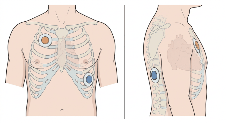
- Position the pads. Anterolateral: one pad parasternally on the right below the clavicle, the other in the anterior to mid-axillary line at the level of the cardiac apex, below and lateral to the left nipple. Anteroposterior: one pad anteriorly over the left praecordium, the other on the back between the left scapula and the spine. The two positions are equivalent in studies of atrial fibrillation; the anteroposterior position is preferred in patients with an implanted device, in the markedly obese and after repeated unsuccessful attempts.
- Connect the ECG cables from the defibrillator to the patient — on most machines the synchronisation reads the cable lead, not the pads.
- Choose a lead with a tall, unambiguous R wave. Increase the amplitude if necessary.
- Press SYNC and check on the screen that a marker sits on every R wave and nowhere else. Without confirmed markers no shock is delivered.
- Preoxygenate and sedate. Wait until the patient does not respond to speech.
- Select the energy (see the table). Charge.
- Call out loudly and check visually that nobody is in contact with the patient or the bed. Pay particular attention to whoever is holding the mask.
- Hold the button down until the shock is delivered — in synchronised mode it comes with a delay, up to one heartbeat later. If you release too early, no shock is delivered.
- Check the rhythm. If a further attempt is made, SYNC must be activated again — most defibrillators revert automatically to unsynchronised mode after every delivered shock.
- If cardioversion fails, step up the energy, change the pad position, press the pads more firmly against the chest, or deliver the shock during expiration. More than three or four attempts is rarely worthwhile.
- Record a new 12-lead ECG once sinus rhythm has been achieved.
Energy levels, biphasic defibrillator
| Arrhythmia | Starting energy | Comment |
|---|---|---|
| Atrial fibrillation | 120–150 J, stepped up | Swedish practice often starts directly at 150–200 J, particularly with a longer duration and a high body weight |
| Atrial flutter | 70–120 J | Often converts at low energy |
| SVT (atrial tachycardia, AVNRT, AVRT) | 70–120 J | Adenosine first in the stable patient |
| Monomorphic VT with a pulse | 120–150 J | Synchronised |
| Polymorphic VT, ventricular fibrillation | Maximum energy according to the manufacturer | Unsynchronised |
Energy scales differ between manufacturers. Follow the energy sequence printed on the defibrillator you are using, not a figure from memory.
Pacemaker and ICD
- Place the pads anteroposteriorly and at least 8 cm from the device so that the current path does not run through the generator or the lead system.
- In ICD recipients, overdrive pacing or internal cardioversion via the device may be an alternative in atrial arrhythmia; discuss this with the arrhythmia unit.
- Interrogate the device after cardioversion — sensing, pacing thresholds and lead impedances — before the patient goes home. Shock-induced threshold rise and loss of programming do occur.
- Be prepared for a pacemaker-dependent patient in whom pacing fails immediately after the shock; have transcutaneous pacing to hand.
Complications
| Complication | Comment |
|---|---|
| Thromboembolism and stroke | The risk is greatest with inadequate anticoagulation; it may occur up to a few weeks afterwards |
| Ventricular fibrillation | Almost exclusively after an unsynchronised shock, in digitalis toxicity or in hypokalaemia |
| Bradycardia, asystole, sinus arrest | Commonest in marked sinus node dysfunction; have atropine and pacing available |
| Skin erythema and superficial burns | The commonest; prevented by good skin contact and dry, shaved skin |
| Sedation-related respiratory depression, hypotension and aspiration | The commonest acute event during the procedure |
| Pulmonary oedema | Rare, described after cardioversion of long-standing fibrillation |
| Myocardial injury with a slight troponin rise | Usually of no clinical significance |
| Damage to a pacemaker or ICD | Avoided by the correct pad distance; interrogate the device afterwards |
Aftercare and follow-up
- Monitoring with ECG, saturation and blood pressure until the patient is fully awake and haemodynamically stable, at least one to two hours.
- A 12-lead ECG about one hour after cardioversion — late recurrences are common and are otherwise not detected.
- A neurological check compared with the baseline status before discharge.
- Inspect the skin under the pads.
- An anticoagulant for at least four weeks, no matter how briefly the fibrillation lasted or how well sinus rhythm is holding. Continued treatment thereafter is governed by the CHA₂DS₂-VA score.
- No driving for 24 hours after sedation, and the patient must not go home alone.
- Plan antiarrhythmic prophylaxis, rate control and investigation of underlying causes — hyperthyroidism, hypertension, sleep apnoea, alcohol, valvular disease — otherwise the arrhythmia will return.
- Explain that recurrence is common and what the patient should do if symptoms occur.
Common pitfalls
- Not checking the sync markers on the screen before every single shock. Sync is switched off automatically after a delivered shock on most machines.
- Leaving sync switched on in polymorphic VT or ventricular fibrillation — the machine then delivers no shock at all.
- Releasing the shock button too early. A synchronised shock is delivered with a delay, not immediately.
- Trusting the anticoagulation history without asking about missed doses. A missed week of DOAC does not appear in the record.
- Forgetting the anticoagulant for the four weeks afterwards because the fibrillation was short-lived or the cardioversion went well.
- Cardioverting with uncorrected hypokalaemia or in digitalis toxicity.
- A pad placed over the device in a pacemaker or ICD recipient, and never interrogating the device afterwards.
- Moist, sweaty or untreated markedly hairy skin — this causes high impedance, failed cardioversion and burns.
- Inadequate sedation. The patient remembers the shock and refuses the next time.
- Charging again and again instead of changing the pad position or reconsidering the indication.


